Chapter 3 – Homeostasis
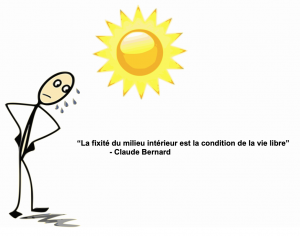
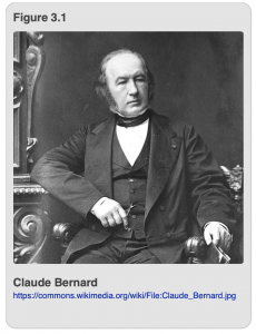
We begin this chapter with a quote from the famous 19th century physiologist, Claude Bernard. Bernard (shown in Figure 3.1) was famous for many things, including several successful attempts to bring science into the field of medicine in preference to its status as an art. He successfully deduced the role of the pancreas in mammalian digestion as well as several aspects of what the liver does. Unfortunately, he accomplished much of this work by performing vivisection on animals, a practice that brought him a fair amount notoriety and scorn from, among others, his wife who ultimately divorced him as a result. If one were to summarize Bernard’s contributions in a phrase, it would be that which appears in the title of Chapter 3 of this book “La fixité du milieu intérieur est la condition de la vie libre.” Translated from the French, it reads: “The fixity of the internal body is the condition of a free and independent life.” We now use the word homeostasis (homeo = similar to; stasis =standing still) to encapsulate the idea that processes inside an organism must be maintained within certain ranges and that there are processes and procedures designed to accomplish just that irrespective of conditions outside the organism.
It is important for parts of the body to maintain critical functions within certain defined limits so that the rest of the body can function properly. This is what is meant by homeostasis, or steady state. For example, in most homes or apartments there is a thermostat that measures the ambient temperature. A typical range for the thermostat in the winter to be set is around 68-70°F. When the room temperature drops below 68°F, the thermostat sends a signal to the furnace to kick on and produce heat. The furnace will continue to do so until the room temperature climbs above 70°F, at which point the thermostat will signal the furnace to shut off. As long as all the equipment is working properly, the room should maintain a comfortable temperature that averages 68-70°F.
The human body works in roughly the same way. Internal detectors measure when the body is too hot, and execute processes that will reduce body heat when the temperature exceeds 98.6°F. Sometimes, it is something as simple and voluntary as taking a rest from activity, or it may be involuntary, like sweating; in either case, both actions will reduce internal body temperature. Similarly, if the body temperature is too low, the brain will send a signal to the body to engage in what physiologists refer to as “futile cycling.” In this process, glucose, an energy-rich sugar, is produced from other energy stores and subsequently destroyed, the net result of which is the generation of heat; the external sign that this is happening is an uncontrollable shaking movement, often referred to as shivering. Using these mechanisms, it is possible to keep the human body pretty close to 98.6°F most of the time.
Temperature regulation is very important to the proper functioning of the bodies of mammals and birds because so many processes depend on body temperature being maintained within a specific range. But, of course, it’s not just body temperature that requires constant supervision and adjustment to assure physiologically appropriate limits, or steady states. Many other processes require this sort of control, making it all the more amazing that this can go on in the human body without the owner of that body even being aware that it is occurring. The job is assigned to the peripheral nervous system and the endocrine system, and it will take care of many of these vital functions without our being consciously aware of it.
Insects, too, must regulate their temperatures so that they can keep their body temperature in an optimal range that supports physiological processes. Interestingly, most insects are ectothermic, which is to say that most don’t have a way to generate heat endogenously. Instead, they have to find a warm place and transfer heat from the environment in order to secure a body temperature that is conducive to life. However, there are some exceptions. Insects that fly generate enormous amounts of heat when flying (due to the exertion of their flight muscles) and may reach a point where they have to dissipate that heat in order to stay within the critical range. One simple option is to stop flying and sit in the shade for a while. But more active approaches to heat loss also occur. For instance, flying insects may have the ability to transfer heat generated in the thorax to the abdomen. The abdomen then serves as a radiator that transfers the heat to the ambient environment. Another creative mechanism for heat transfer occurs in mosquitoes. After taking a hot blood meal and which increases their internal body temperature, female mosquitoes will secrete a drop of liquid from their abdomen. Evaporation of the drop cools the insect, just as the evaporation of sweat cools the human body.
We mention thermoregulation because it is a classical, well-known example of homeostasis. Also because there are clear similarities in humans and insects with respect to the need to regulate body temperature, and clear differences in the mechanisms by which it is done. The kinds of control exerted to maintain body temperature can be generalized to other physiological systems. In fact, the processes of digestion and excretion in both humans and insects repeatedly rely on homeostatic feedback loops and, thus, provide multiple ways to understand this fundamental concept in biology.
The goal of the digestive and excretory systems in both humans and insects is to take food into the body, digest it, and extract nutrients from it to power the rest of the body. In both insects and humans, the basic site of digestion is an alimentary canal, which is the term that describes the whole passage along which food passes from mouth to anus. In other words, the alimentary canal extends nearly the entire length of the body. In order to understand the similarities and differences in humans and insects, it is necessary to study the anatomy of both types of organisms. As we did for the respiratory and circulatory systems in Chapter 2, we will first review the digestive and excretory processes in humans and then discuss them in insects. Similarly, we will point out several of the adaptations in different groups of insects that allow them to survive and thrive by feeding on a variety of food sources.
Human Digestive and Excretory Systems
In humans, the digestive system consists, in essence, of the mouth, esophagus, stomach, and intestines (Figure 3.2). The companion excretory system is comprised of the intestines, rectum, and anus that handle solid waste; and the kidneys, ureters, bladder, and urethra that process water-soluble waste. The action all begins in the mouth where food is ingested, and chewed if it is solid. In addition to the mastication process, where food is physically split into smaller pieces, the chewed food is treated with the enzyme, amylase, in the saliva, to begin chemically breaking down complex sugars into their simpler forms. Then the food is swallowed and passes through a long tube called the esophagus to get to the stomach. Muscular contractions in the stomach, along with highly acidic digestive juices and enzymes, further break down the food (proteins into amino acids; starches/complex carbohydrates into simple sugars; fats into lipids). The bolus of food then passes into the small and large intestines. The small intestine is quite long and is lined with special cells that absorb nutrients from the processed food. These cells are also in close contact with capillaries, allowing the small intestines to shunt nutrients into the bloodstream so that the nutrients can then be distributed to all of the cells in the body.
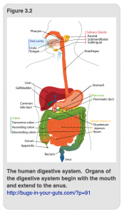
After as many nutrients have been extracted from the solid food as possible, the remaining undigested material is then sent to the large intestine (also called the colon), where water and salts are extracted from the solid waste. The large intestine and rectum then return valuable salts and water to the bloodstream via capillaries. The remaining material cannot be further digested or recycled and is therefore eliminated from the anus as solid waste (feces).
The preceding description is a very cursory analysis of how digestion occurs in humans. There are many other processes and organs that one could discuss in this context. For instance, there are some ancillary digestive organs such as the liver, gallbladder, and pancreas that supply hormones and/or enzymes that aid in food processing and nutrient absorption. Further, if something toxic is ingested or a toxic byproduct is released during the digestion process, it can be sent to the liver to be detoxified or rendered harmless. Humans also have a vestigial organ (an organ that was necessary in our ancestors but is now nonfunctional) called the appendix which serves no function in digestion and appears to be an evolutionary relic whose sole function, if you can call it that, is to become inflamed causing a life-threatening infection. Alas, it is not a perfect system.
The liquid foods that you ingest (i.e. soups, smoothies, and beverages) also travel down your esophagus, to your stomach, through your small intestine where the nutrients are extracted and distributed into the blood, and then to your large intestine where most of the water is absorbed. However, excess water and water-soluble waste products from the food, including the byproducts of protein metabolism, are eliminated using different excretory organs. Here the function of processing and expelling water-soluble wastes, such as urea, are given to the kidneys, ureters, bladder and urethra (Figure 3.3).
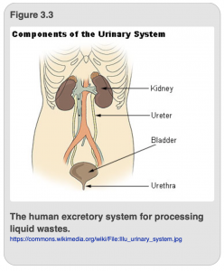
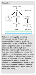
Most humans eat foods such as meat, eggs, cheese, and legumes (beans, peas, and lentils) that are rich in protein, and hence nitrogen (the important element abundant in protein). Our bodies break down these proteins into individual amino acids, and then use these to produce our own proteins and nucleic acids (DNA and RNA). However, we can certainly have too much of a good thing—byproducts of nitrogen metabolism are generally released as either ammonia (NH3), as in the toxic cleaning product, or its ionized form, ammonium (NH4+), both of which are water soluble and highly harmful to our cells. Thankfully, our liver converts these nasty byproducts into a non-toxic, but still water-soluble chemical called urea (Figure 3.4). The liver releases urea into the bloodstream, where it floats along until it reaches our kidneys. Our kidneys act as our major blood-purification filters, removing urea and other water-soluble waste products, as well as returning useful substances, like glucose and amino acids back into the blood stream. After all of the harmful substances have been removed and the useful substances have been recycled into the bloodstream, the remaining liquid waste moves from the kidney through the ureters to the bladder, where it is stored temporarily before being voided through the urethra as liquid urine.
In addition to their pivotal role in water-soluble waste removal, the kidneys are also directly responsible for various homeostatic functions that include the regulation of blood pressure through the maintenance of salt and water balance, and the acid-base balance in our cells. Our kidneys also release hormones that regulate our blood pressure, control the production of red blood cells, and produce an active form of vitamin D that keeps our bones healthy and strong. So, if you haven’t yet, stop and thank your kidneys for all that they do for you!
Insect Digestive and Excretory Systems
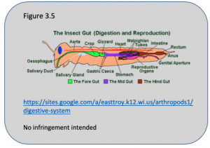
The function of the digestive system in insects is remarkably similar to that in humans. However, the structure is somewhat different. Functionally, the insect digestive system is divided into three parts: the foregut, midgut, and hindgut. Components of the foregut include the mouth, esophagus, crop, and gizzard/proventriculus (when it is present). Similar to human teeth and tongues, the insect mouthparts are used to prehend (hold on to) food, crush it into smaller pieces, and mix it with saliva for preliminary digestion before it moves to through the esophagus. In several insect species, part of the esophagus has been expanded into a specialized organ called the crop which serves as the main holding area for food (Figure 3.5). The crop empties into the gizzard/proventriculus, which physically grinds the food into smaller pieces, much like the muscular contractions of our stomach, making the food easier to digest by chemical means.
After the food leaves the gizzard/proventriculus, it enters the midgut, which consists of stomach/ventriculus and gastric caecae (when present). Active digestion and nutrient absorption take place in the stomach as well as the gastric caecae. The gastric caeca are finger-like projections that mark the transition between foregut and midgut. In addition to providing enzymes and other aids to digestion, the gastric caecae expand the surface area available for the transfer of nutrients (the products of digestion) into the hemolymph. The gastric caecae may also harbor protozoa (eukaryotic, single-celled organisms), bacteria, and/or archaea that can assist with digestion.
After processing by the midgut, the food bolus moves into the hindgut. The transition from the mid- to hind-gut is marked by the presence of Malpighian tubules, which are spaghetti-like projections that have a sealed end that floats in the body cavity and an open end that empties directly into the hindgut. The major excretory organs and functions are also found in the hindgut (similar to humans!). This is the site of the rectum and its affiliated rectal pads absorb water. Similarly the anus is the site through which solid waste is expelled.
Just like humans, insects have to detoxify nitrogenous waste products (i.e. NH3 and NH4+). However, rather than converting these into water-soluble urea, the fat body convert ammonia to water-insoluble uric acid, which the Malphgian tubules then filter out of the bloodstream. (Note that this is similar to how our liver detoxifies ammonia into urea and our kidneys filter it out and remove it.) The Malpighian tubules further help insects conserve water by reabsorbing salts and some water from partially digested food and returning them to the hemolymph circulation. After processing by the Malpighian tubules, the digested material passes over the rectal pads where even more water is extracted prior to elimination. Therefore, for most terrestrial insects, they excrete a dry, hard fecal pellet.
After all is said and done, material in the insect digestive tract that cannot be digested or recycled will pass through the anus and be eliminated as frass—the correct insect-based term for plant-derived solid waste. As you can tell, there are many similarities in the digestive and excretory processes in humans and insects. There are also many differences that arise, in part, because of the diversity of food stuffs that insects eat as well as the different environments that insects inhabit.
Diversity in Insect Digestive and Excretory Systems
The excretory system has two basic functions in both humans and insects: excrete nitrogenous wastes and maintain water balance and homeostasis. Humans are decidedly terrestrial. However, some insects have one or more life stages live in an aquatic environment. In either case, the maintenance of homeostasis is a primary concern. Organisms will face unique digestive and excretory challenges depending on both their chosen environment (i.e. terrestrial vs. aquatic) and the food sources that they consume (i.e. solid plant material vs. nectar vs. other insects).
As a general consideration, organisms in a terrestrial environment will want to conserve water and excrete salts. This is especially true for terrestrial insects, as they are especially susceptible to dehydration. The small size of insects creates a relatively large surface area relative to their volume. This means that insects have proportionally more of their body exposed to the environment. Therefore, terrestrial insects have acquired several unique mechanisms, such as the ones that we have already discussed in this chapter (excreting nitrogenous waste as water-insoluble urea, reabsorbing water in their Malpighian tubules and rectal pads) as well as others, such as only opening their spiracles intermittently when they breathe (discussed in Chapter 2), and covering their exoskeletons with wax (to be covered in Chapter 4) to help them conserve as much water as possible.
In contrast, those organisms that live in an aquatic environment will want to retain salts and eliminate excess water. For this reason, most aquatic insects excrete their nitrogenous waste directly as ammonia (NH3), and many have specialized structures, such as rectal gills (dragonflies) or anal papillae (mosquito larvae), that allow them to uptake salts from their aquatic environments. Insects that have both aquatic and terrestrial phases at different stages of their life cycle have some especially interesting challenges, and their excretory systems change as they develop, allowing them to meet these challenges.
As you might expect, there is a lot of variation among insect digestive systems according to what they eat. What are the kinds of factors that might dictate alterations in different parts of the digestive system? We have already broached this question in an observation made earlier in this chapter when we noted that insects might have chewing, biting, or sucking mouthparts depending on the type of food they eat. Fortunately, this is one of those moments when the form of a structure gives you an idea of its function that we mentioned in the introduction of the book. As you can see from Figure 3.6, there are obvious differences in mouthparts that closely correspond to the insect’s preferred food source. For example, Figure 3.6A shows the chewing mouthparts of a grasshopper. Grasshoppers have large, hard mandibles with teeth (shown in green) that they uses to grind up food, as well as finger-like palps on its maxillae (yellow) and labium (blue) that they uses to hold and manipulate the food. Several diverse groups of insects have chewing mouthparts that share basically the same structure to those of the grasshopper, providing strong evidence that this is the ancestral way of feeding in most insects.
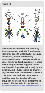
However, as insects evolved and adapted to take advantage of new food sources, their mouthparts were modified in unique ways. For example, Figure 3.6B shows the mouthparts of a honey bee. Not only does she have specific adaptations to do work around the hive, like molding wax with her small mandibles, she also has a tube-like mouth (comprised of the maxillae and labium) that she can use to suck nectar from plants. Next, Figure 3.6C shows the mouthparts of a butterfly, which have been modified into a very long tube called a proboscis. The proboscis (formed from the base of the maxillae) is so long it has to be rolled up when it is not in use. When a butterfly finds a flower that is its food source, it will unroll the proboscis and insert it into the flower to withdraw nectar. Finally, Figure 3.6D shows the mouthparts of an adult female mosquito. Here, the majority of the mouthparts (mandibles, maxillae, and labrum) have all become long and thin and wrapped together to form a stylet that is protected in a sheath formed from the labium. When a female mosquito blood-feeds, the sheath bends back and the mosquito is able to insert her thin stylet into our skin to probe and eventually pierce an individual capillary and draw out our blood (Video 3.1).
In addition to requiring different mouthparts to ingest various food sources, additional anatomical and physiological modifications to the digestive tract are also required. As you can see from Figure 3.7, it is possible to tell just by looking at the relative sizes and shapes of components of an insect’s alimentary canal what it eats. There is, perhaps, a bit more detail in this diagram than would be optimal for a non-entomology audience, but nonetheless, it shows you very quickly that insects that eat different foods have guts that have been modified in different ways. The diagram on the left (Figure 3.7A) shows you the digestive tract of a springtail. These are very primitive hexapods (not considered to be true insects), which eat decaying vegetable-matter, fungi, and bacteria. Their guts are little more than a simple tube and lack most of the structures (i.e. gastric caecae, Malpighian tubules, etc.) that characterize other insect guts. The second schematic (Figure 3.7B)shows the alimentary canal of a grasshopper. Although grasshoppers are primarily plant eaters, they will also scavenge and consume the bodies of other dead arthropods. They digest complex plant carbohydrates mainly in their well-developed crops, and process protein and fats in their gastric caeca. They also have numerous Malpighian tubules to process and remove nitrogenous waste. Caterpillars (Figure 3.7C) feed exclusively on plants, and hence have greatly expanded salivary glands to promote initial digestion and processing of food, as well as large midguts where the complex plant carbohydrates can be broken down and absorbed.
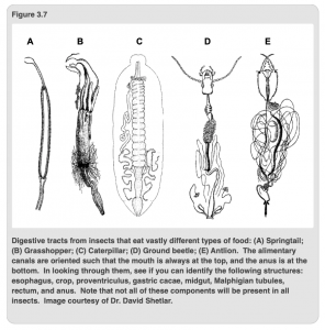
As the nitrogen content of most plants is relatively low, caterpillars typically have fewer Malpighian tubules. This is in contrast to predatory insects, like the ground beetle and antlions, which subsist on high-nitrogen diets. Ground beetles (Figure 3.7D) eat their prey by first breaking them apart with their mandibles. The chunks of arthropod goodness are further broken apart in their well-defined gizzard/proventriculus before reaching the gastric caeca where the bulk of protein digestion occurs. In contrast, antlions (Figure 3.7E) have long, sharp hollow mandibles which they use to suck out the liquid juices of their preferred food source: ants. Nutrients are absorbed quickly in their expanded midguts, and the excess nitrogenous wastes are processed by their long and numerous Malpighian tubules.
Chapter Summary
We realize that we have introduced a lot of terminology and conceptual knowledge in this chapter. Our intent is not to overwhelm you, but to give you a basic understanding of how humans and insects ingest food, break it down into usable components, and excrete the waste products in order to maintain homeostasis. In addition to this, there are a couple of other fundamental concepts that should now be clear.
First, all environments present a range of challenges to organisms that inhabit them. We have looked in detail at the different challenges besetting humans and generic terrestrial and aquatic insects. We hope that you are now familiar with how insects and humans experience the processes of digestion and excretion are able to identify ways in which they are similar and ways in which they differ.
Second, because insects occupy so many different environments and may eat vastly different things, you should be able to identify key ways in which the processes of digestion and excretion vary in insects according to their feeding habits and environments. For example, how do you expect the mouthparts and alimentary canal of a true bug that feeds on plant juices to differ from those of a caterpillar that ingests chunks of leaves? Similarly, how might a terrestrial insect predator, such as a ground beetle, differ in both its alimentary canal and method of nitrogenous waste excretion from an aquatic dragonfly nymph, which is also predatory? When considering these questions, you should be able to see and articulate what is meant by the idea the function follows form in insects and humans. Indeed, the tight relationship between form and function is a unifying theme in biology, and hence, you will see this theme repeated throughout the book.