Skill Group: Dermatology
Goal Four: Trichogram
Before You Start …
Make sure you have the following equipment: Clean glass slides, mosquito hemostats, or rubber covered hemostats, fingers, cover slips, mineral oil, pencil, microscope
Introduction
Plucking hairs from the skin and examining them under the microscope is helpful for examining the hair shaft and bulb. These are useful in diagnosing self-inflicted alopecia, dermatophytosis, Demodex, Cheyletiella ova, color dilution alopecia, nutritional or congenital hair dysplasia, anagen and telogen effluvium, endocrine alopecia and pigmentary defects as well as assessing hair growth phase.
Learning Outcomes
By the end of this lesson, you should be able to:
- Collect, examine and interpret the results of the trichogram
- Explain how to use a trichogram to evaluate a pet for parasitic and congenital alopecic diseases.
- Interpret a trichogram to determine if a pet is pruritic or not.
- Describe the difference and significance between an anagen and telogen hair bulbs.
Technique
Trichography is performed using either fingers (if infectious etiologies are not expected), however, using rubber covered hemostats or mosquito hemostats makes for acquiring a more precise sample. These are easily performed at most sites.
Here are the steps for performing this technique; a video that walks through these steps is available:
- Label a glass slide on the frosted area with the patient’s name and area of sample collection for example: “Gypsy” left lateral thorax.
- Place a large drop of mineral oil in the center of the labeled slide.
- Identify the affected site. Grasp a small number of hairs at their base where they enter the skin. Do not pinch the skin with your fingers or hemostats.
- Gently but forcibly pull the hairs out of the follicles in the direction that the hair are growing. This will epilate the hairs from the follicle and minimize traumatic artifacts
- Place the sample in the mineral oil on the slide ensuring all hairs are lying in the same orientation.
- Place a coverslip over the sample and examine with the microscope under 4X- 10X objective.
Interpretation
Hair Tips
Hair tips are evaluated to determine if the patient is pruritic (especially cats) or if the cause of the hair loss is non-traumatic (e.g. endocrine disease, follicular dysplasia). Pruritic animals break the tips off the hairs, leaving a broken end that can be detected early. This determination is especially useful in feline patients when owners are not convinced that the patient is pruritic because of its secretive nature.
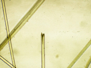
Hair Roots
Hairs will either have an anagen or telogen root (bulb). Anagen bulbs are rounded and smooth and often pigmented and soft so the root may bend. Telogen bulbs are club or spear- shaped, rough surfaced, nonpigmented, and generally straight. Normal adult animals have an admixture of anagen and telogen hairs, the ratio of which varies with breed, season, management and numerous other factors. Normal anagen:telogen ratios have not been established for all breeds of animals. No normal animal should have all of its hairs in telogen, this would indicate telogen defluxion, or possibly nutritional, endocrine or metabolic diseases. Poodles have prolonged growth periods where most hairs are in anagen, with relatively few hairs in the telogen stage.
Hair Shafts
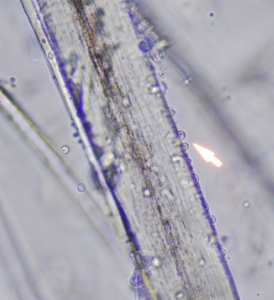
A normal hair shaft is uniform in diameter and tapers gently to the tip. Curly or wavy-coated animals have twisted hair shafts. Hair pigmentation depends on the coat color and breed of animal, but should not vary greatly from one hair to the next in regions where the coat color is the same.
Hair shafts can have pigment clumping that leads to breakage; this is seen with color dilution alopecia.
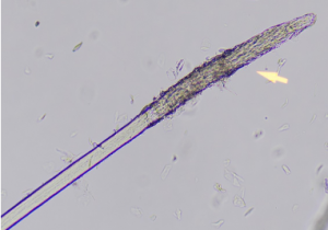
Hair shafts can appear swollen, damaged and have a fuzzy cortex with ends that are frayed (like a broom).
With closer examination the damaged area may reveal small spherical structures called ectothrix spores of dermatophytes.
Hair casts are seen in diseases associated with follicular keratinization abnormalities, such as sebaceous adenitis, follicular dysplasia, zinc responsive dermatosis, demodicosis and endocrinopathies.
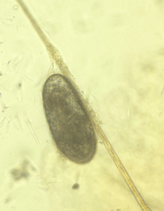
Finally, ectoparasite eggs attached to the hair shaft may be visible with pediculosis and cheyletiellosis.
Wrapping Up
You are now able to perform a trichogram. You now understand when to perform this test to help you confirm dermatologic disease. You are now able to recall expected abnormalities in the hair associated with disease. These skills will help you formulate differential diagnoses.
Next, we will learn how to perform fungal examination and culture.
Before Moving On …
Use the self-check activities below to check your understanding.
(Under development)