Skill Group: Dermatology
Goal One: Dermatology and Otic Examination – Examination of the Skin
Before You Start …
Make sure you have the following equipment: Examination table, ceiling or wall mounted LED examination lights, gloves, lab coat and/or scrubs, ± handheld 5X-7X LED medical magnifying glass
Introduction
A through dermatological evaluation is difficult to achieve with the owner providing restraint (and also providing distraction from the business at hand). Another barrier to performing a through dermatological examination is the time constraint placed on general practice appointments. Consider scheduling the patient’s initial dermatology examination as a special procedure appointment so enough time has been set-aside to do a very thorough dermatological evaluation and a minimum database.
Learning Outcomes
By the end of this lesson, you should be able to:
- Prepare your PPE and the room to ensure an excellent dermatological examination can occur.
- Educate the assistant on how best to help during the dermatological examination.
- Demonstrate how to perform a methodical dermatological examination.
- Identify primary and secondary lesions.
Technique
Thorough dermatologic examination is performed by placing the pet on an examination table with proper gentle restraint. Uncooperative patients should be sedated as required to allow a complete and through examination in every case.
Here are the steps for performing this technique; a video that walks through these steps is available:
- Ensure adequate lighting.
- Put your examination gloves on.
- Perform a general physical examination. The skin is often an indicator of systemic disease that the owner may not appreciate.
- Start with examining the entire animal from a distance to get an impression of its overall health, its attitude, obvious abnormalities and the distribution of the disease. The distribution of lesions often helps in prioritizing differential diagnoses.
- The veterinary assistant should provide gentle restraint of the pet on the side of the table opposite of the examiner.
- After an impression is obtained from a distance, the skin should be examined more closely and palpated.
- Examine the skin and hair coat thoroughly in a systematic fashion. For example, always start at the nose and work backwards, first with the pet upright and then again with the pet restrained in lateral recumbency to examine the ventrum.
- Inspect all visible mucous membranes as they are encountered.
- Examine every centimeter of skin. Thick-coated animals may need to be clipped so that the skin and skin lesions can be visualized adequately.
- What is the texture of the skin (rough, smooth)?
- Is the skin the proper thickness for the body location?
- Is the skin the appropriate color considering the breed and hair coat color?
- Check the elasticity (does it snap back into place?)
- Does the skin feel excessively warm or cold?
- Examine the hair coat
- What is the texture of the hair (fine, course, dry or oily)?
- Does it epilate easily?
- Is their partial hair loss (hypotrichosis) or complete alopecia in areas where it is normally present?
- Is the hair loss diffuse or are there multiple focal areas of alopecia?
- At regular intervals, the hair should be parted or skin examined by rolling it up in folds to separate the hair in short-coated dogs.
- Identify primary lesions.
- Identify secondary lesions.
- Examine the interdigital spaces, ungual folds, claws and footpads while the animal is retrained in lateral recumbency. If the pet will not lie comfortably in lateral recumbancey, then examine these areas on the paws in the standing patient.
- Record your findings in the medical record.
| Primary Lesion | Image | Description |
|---|---|---|
| Pustule | 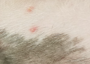 |
|
| Nodule | 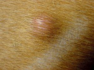 |
|
| Vesicle | 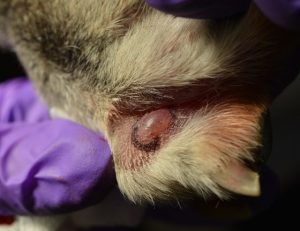 |
|
| Tumor | 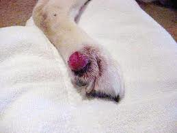 |
|
| Papule | 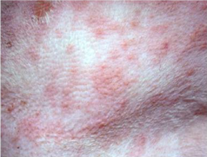 |
|
| Wheal | 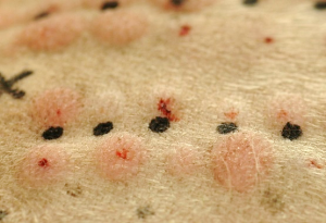 |
|
| Patch | 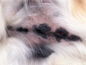 |
|
| Plaque | 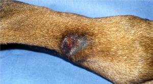 |
|
Wrapping Up
You have learned how to position the patient and instruct the staff on proper restraint so a through dermatologic examination can be performed and primary lesions identified. These skills are essential for formulating differential diagnoses and determining necessary diagnostic tests.
Next, we will learn how to perform an examination of the ears.
Before Moving On …
Use the self-check activities below to practice identifying lesions.