Skill Group: Clinical Skills
Dental Anatomy
Introduction
Understanding what structures are considered a portion of the tooth itself vs. a supporting anatomical structure is important, as it helps us to not only identify disease in the mouth, but also enhances our grasp of extraction principles.
In the previous lesson, you learned how to identify each tooth using the modified Triadan system, as well as the importance of understanding dental formulas. We will now look at the structures of the teeth and the supporting periodontal structures, and we’ll also learn how to identify the location of areas on these structures using directional terminology.
Learning Outcomes
By the end of this lesson, you should be able to:
- Correctly identify the tooth structures vs periodontal structures.
- Correctly identify the function of each structure (tooth and periodontal).
- Locate a lesion using two directional terms.
The Categories of Dental Anatomy
Dental anatomy is broken down into 2 categories:
- Structures of the tooth. These structures make up the substance of the tooth itself.
- Structures of the periodontal attachment apparatus, or periodontium. Each of these structures helps to anchor the tooth into the mouth, while minimizing damage to the animal when the tooth is performing its function.
Understanding the structure and function of each part of the tooth helps us to understand what treatments we may need to perform if we found abnormalities while charting the oral structures.
Tip: As you review the descriptions below, reference the “Medical illustration of a maxillary canine in a dog” figure in Disorders of Dental Hard Tissues in Dogs (Niemiec, 2014).
Structures of the Tooth
Enamel:
- Covers the crown of the tooth, which is visible above the gum line.
- Hardest and most dense tissue in the body.
- Contains the highest % of minerals, 96% hydroxyapatite in which the primary mineral is crystalline calcium phosphate; remaining 4% is water and fibrous organic material.
- Protects the tooth from damage during daily use, potentially painful temperatures or chemicals.
- Unable to regenerate.
- Translucent.
Cementum:
- Covers the root of the tooth, which is below the gum line.
- Similar to bone in structure.
- Vital, able to repair to a limited degree.
- Anchors the periodontal ligament, which attaches teeth to the alveolar bone.
- Harder than bone, but softer than enamel and dentin.
Dentin / Dentine (either is correct):
- Covered by enamel on the crown and cementum on the root.
- Surrounds the entire pulp.
- Yellow in color and porous in nature, greatly affects the color of the tooth due to the translucency of the enamel.
- Harder than bone, but softer than enamel.
- 3 types of dentin:
- Primary – forms before eruption.
- Secondary – forms after eruption, continues to be deposited as animal ages.
- Tertiary – reparative, disorganized, forms after trauma to odontoblasts.
Pulp:
- Located in the center of the tooth.
- Consists of blood vessels, lymphatics vessels, nerves, fibroblasts, collagen fibers, odontoblasts, and many other cell lines.
- Supplies oxygen and nutrients to local cells and provides exit route for metabolic waste.
- Part of the pulp-dentin complex, called endodontium.
- Gets smaller with age due to the continued deposition of dentin.
Structures of the Periodontium
Gingiva (gums):
- Mucosal tissue in the mouth surrounding the teeth and covering the bones of the jaw.
- Stratified squamous epithelium with some keratinization.
- Forms a seal around the teeth for protection and stabilization in the bone.
- Only visible structure of the periodontium.
- 2 types of gingiva:
- Free gingiva: It is not directly attached to the tooth, and it forms a groove called the gingival sulcus. The gingival sulcus is where we insert our dental probe to measure depth; depth of greater than 3mm in dogs and 0.5-1 mm in cats is considered ABNORMAL.
- Attached gingiva: It is directly attached to the cementum and alveolar bone, making it immovable, firm. and resilient.
Cementum: (see above) While Cementum is considered a part of the tooth itself, it is also critical to the periodontal attachment apparatus.
Periodontal Ligament:
- Strong fibrous tissue connecting the cementum of the tooth to the alveolar bone.
- Contains connective tissue, collagen, nerves and blood vessels.
- Commonly abbreviated PDL.
- Functions as shock absorber for chewing.
- Force necessary for chewing is regulated by the nerves of the PDL.
Alveolar Bone:
- The part of the mandible and maxilla that surround and support the alveolus or socket of the tooth.
- Provides structural support for the root of the tooth.
- Contains 4 layers – periosteum, dense compact bone, cancellous bone, and the cribriform plate (also known as the Lamina Dura).
- The Lamina Dura lines the alveolus and is the attachment site for the PDL, which then attaches to the Cementum to hold the tooth in place.
Anatomical Directional Terms
The anatomic directional terms in the mouth are slightly different than other places in the body; in the mouth, we identify our location in reference to the tooth and the median line:
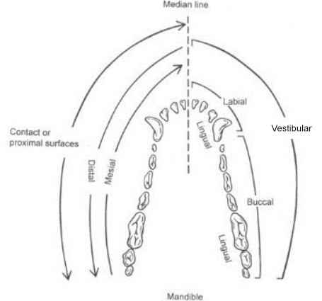
- Toward the median line is called mesial; and
- Away from the median line is called distal.
If we are locating lesion in the oral cavity, we use two terms to identify our location:
- First is the term in reference to the tooth and the medial line, as explained above – mesial or distal;
- Second is which side of the tooth the lesion has occurred:
- Buccal lesions occur on the side of the tooth closest to the cheek;
- Labial lesions occur on side of the tooth closest to the lip (typically located on incisors);
- Palatal lesions occur on the side of the tooth closest to the palate in the maxilla; and
- Lingual lesions occur on the side of the tooth closest to the tongue in the mandible.
Practice Time!
Wrapping Up
In this lesson, you learned the structures of the teeth and the supporting periodontal structures, as well as how to identify the location of areas on these structures using directional terminology. In the next lesson, you’ll learn about how to distinguish between normal and abnormal dental findings.
Before Moving On …
Use the self-check activity below to practice identifying locations on the tooth using directional terms.