Skill Group: Dermatology
Goal Six: Bacterial Culture
Before You Start …
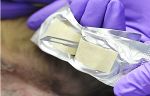
Make sure you have the following equipment: sterile 25 G needles, sterile #10 blade, culturette specimen swabs in transport media; sterile instruments and gloves, sterile punch biopsy.
Introduction
Bacterial culture and sensitivity are used to identify potential pathogenic organisms and guide the selection of appropriate antimicrobial therapy. When cytologic examination reveals rod-shaped organisms, or when cocci are seen but appropriate empirical therapy is ineffective, bacterial culture and susceptibility testing are indicated (see table for indications of when to perform a culture and sensitivity).
Learning Outcomes
By the end of this lesson, you should be able to:
- Identify sites for bacterial culture, collect samples for the culture and sensitivity testing.
- Outline the different techniques used to perform a bacterial culture from a pustule, papule, crust and epidermal collarette.
- Describe criteria that mandate a bacterial culture be performed.
- Define lesions that should not be sampled for bacterial culture.
Technique
Empirical Antibiotic Therapy is appropriate when all of the following apply:
- Non-life-threatening infection.
- First episode of a skin infection.
- Clinical lesions are consistent with a superficial pyoderma.
- Cytology is consistent with a staphylococcal infection.
- No reason to suspect antibiotic resistance.
Bacterial culture & sensitivity are necessary when any of the following apply:
- Life-threating infections, as the first-choice antibiotic must be effective.
- Clinical lesions are consistent with a deep pyoderma.
- The clinical signs and cytology are not consistent with each other.
- Rod-shaped bacteria are seen on cytology, as their antibiotic sensitivity is notpredictable and may be limited.
- Where antibiotic resistance is more likely:
- After one or more broad-spectrum antibiotic courses.
- Non-healing wounds.
- Postoperative and other nosocomial infections.
- The owner or animal has recent healthcare contacts.
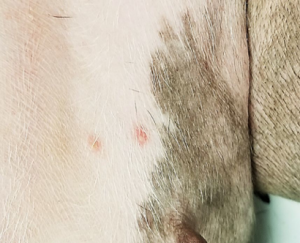
Bacterial culture is performed from lesions that represent a potential bacterial infection. There are several techniques for collecting samples for bacterial culture of the skin, and the type of method often depends on the lesions available for sampling.
- Selection of appropriate lesions for culturing is critical (ideally primary lesions).
- It can be useful to take several samples if there are multiple lesions, especially if cytology findings differ.
- Moist erosions and many crusts may be contaminated by bacterial overgrowth.
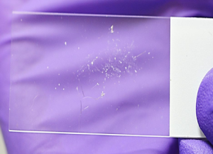
- After sampling lesions, always follow-up with collecting a sample for cytology from the lesion to ensure that the sample submitted has a high likelihood that it has
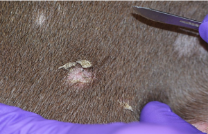
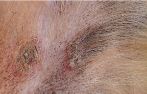
Sampling Lesions that Represent Superficial Bacterial Folliculitis
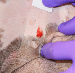
Pustule
Best lesion to sample. Do not sterilize or wipe the lesion with disinfectant.
- Using a sterile 25G needle, lance the intact pustule.
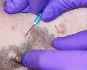
- Touch a sterile culturette to the purulent discharge then place in transport medium for transport to the lab.
- Alternatively, the purulent material on the tip of the needle may be transferred to the tip of the culturette swab.
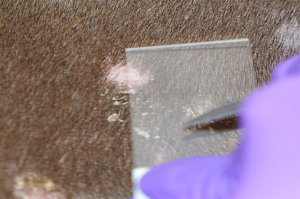
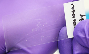
- Collect cytology by performing an impression smear of the site.
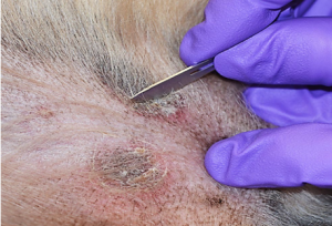
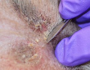
Crust
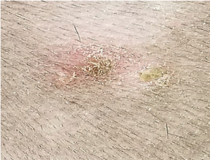
Do not sterilize or wipe the lesion with disinfectant.
- Using either a sterile needle, a sterile #10 blade or mosquito forceps (wipe the forceps with alcohol before), reflect or elevate the crust off the lesion.
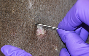
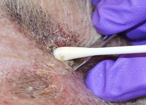
- Touch a sterile culturette to the purulent discharge or to the moist exposed skin surface under the removed crust.

- Perform cytology of the lesion after the sample for culture has been collected.
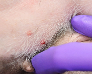
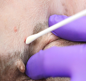
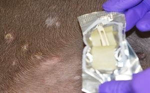
Epidermal collarette
Do not sterilize or wipe the lesion with disinfectant.
- Using a sterile blade or needle, elevate the leading edge of the stratum corneum (i.e. the collarettes).
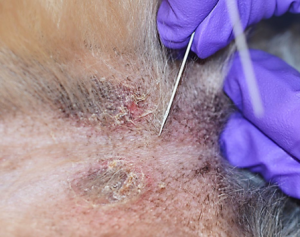
- Roll a sterile culturette on the moist exposed skin surface under the stratum corneum.
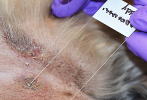
- Place sample in transport medium for transport to the lab.
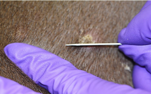
Papule
A tissue sample collected by punch biopsy is necessary.
- After administering local anesthesia (see next section on Skin Biopsy for Histopathology), wipe the surface of the skin once with an alcohol soaked sterile gauze swab. Allow to dry.
- Using a sterile 3 mm punch and sterile instruments, collect the tissue sample that includes the papule and place the sample in a sterile container for transport to the lab.
- The skin is sutured with either simple interrupted sutures or a cruciate suture.
Sampling Lesions that Represent Deep Pyoderma
Furuncles
Needle aspirates may be taken and cultured.
Plaques, Nodules, Fistulous Tracts
A tissue sample is collected aseptically by skin biopsy following the technique described above for papules with the exception. However, skin surface is disinfected more thoroughly using chlorhexidine scrub and alcohol or Zephiran (benzalkonium chloride), rather than just a single wipe with an alcohol gauze swab.
There is conflicting evidence that local anesthetic may be bactericidal; therefore, a ring block, local nerve block or general anesthesia may be needed to aseptically collect 4-8 mm punch biopsies, or full thickness wedge biopsies for tissue cultures.
Wrapping Up
You are now able to select appropriate lesions to collect a sample for a bacterial skin culture. You know how use an appropriate method for collection based on the lesion type. This skill allows you to confirm your cytological findings, select the correct antimicrobial treatment for your patient or help rule-out an infectious dermatosis.
Next, we will learn how to perform skin biopsy.
Before Moving On …
Use the self-check activities below to check your understanding.
(Under development)