Skill Group: Clinical Skills
Periodontal Disease – Gingivitis and Periodontitis
Dr. Stephen Horvath and Krystal Bowers
Introduction
Periodontal disease (PD) is one of the most common health problems in small companion animals with prevalence reports ranging from 60-100% of patients. As you will learn, details in these studies lead one to appreciate that PD is highly under diagnosed via an awake visual oral examination. This is likely associated with the fact that the majority of this disease process occurs underneath the gum-line, where it is hidden from view. Thus, the presence of dental tartar, or lack thereof, does not always correlate with the degree of PD. Additionally, the importance of oral health’s role in systemic health cannot be understated; the body is connected. There exists a multitude of mis-information and a lack of understanding concerning this connection. Recognizing and understanding this disease and its connection to systemic health will allow you to educate your clients on the importance of good oral health. This knowledge will empower you to recommend annual prophylactic dental care, and to make appropriate diagnostic and therapeutic recommendations for your patients.
Stimulate Prior Learning
You’ve previously learned how normal gross and radiographic dental anatomy, how to systematically perform a probing oral exam (looking for periodontal pockets and furcation exposure), and how to take dental radiographs . Now you will use that knowledge to identify periodontal disease, determine and describe the stage of PD, and to develop an appropriate treatment plan.
We need to understand the dental formulas outlined in Module 2, normal anatomical structures and oral anatomic directional terms as outlined in Module 3 and from the VME 1 anatomy course, and be familiar with dental radiographic anatomy and orientation as outlined in Lesson 6. We will provide additional information necessary in this module as well.
Learning Outcomes
Upon completion, you will be able to understand and recognize PD, Subjectively diagnose PD via visual examination, diagnose the stage of PD on a per tooth basis via dental radiographs, and use this information to make appropriate therapeutic recommendations.
By the end of this module, you should be able to:
- Understand the pathophysiology of periodontal disease (PD) and it’s 2 stages
- Demonstrate an understanding of how to diagnose PD subjectively and objectively with dental radiographs
- Demonstrate an understanding of and an ability to diagnose the given stages of PD
- Demonstrate an understanding of the common methods used to treat periodontal disease (root planning – open and closed, perioceutical treatments, dental extraction)
- Demonstrate an ability to develop an appropriate treatment recommendation for the various stages of periodontal disease
Periodontal Disease- Gingivitis and Periodontitis
Read the following article in Today’s Veterinary Practice to understand the pathophysiology and scope of periodontal disease, and the importance of treating this condition early through prophylactic dental care.
Visual Examination- Subjective Diagnosis of Periodontal Disease
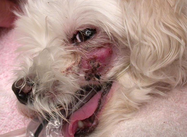
As described below diagnosing a stage of periodontal disease relates to a single tooth. Therefore, patients’ generally have more than one stage present during evaluation. The degree of this variation can dictate how severe the patient’s periodontal disease is and thus how long a given dental procedure may take. The caveat being one needs dental radiographs to fully diagnose each tooth. Therefore, the diagnosis of periodontal disease in an awake patient, during a routine physical exam, encompasses a degree of subjectivity. For this we examine the amount of dental tartar that is present on the crowns, inspect for severity of gingivitis, and for the presence of gingival recession and tooth fractures. We then add all these observations together to generate a subjective severity to the diagnosis of PD. Clinical experience can improve your suspicion, however there are always surprises that can be lurking under the gum-line. Surprises that we don’t fully understand until the patient is anesthetized, and we can perform a full oral exam along with full mouth intra-oral dental radiographs.
Thus, a useful method for documenting your diagnosis of periodontal disease is to use the following subjective qualifying scheme: mild, moderate, or severe, or a combination such as mild-moderate or moderate-severe. Ex: Moderate periodontal disease.
Clinical Pearl – How we manage things in the Community Practice Dental Service:
- When recommending a therapeutic dental cleaning and procedure we must provide the owner with an estimated cost which can be tricky in the face of uncertainty.
- Rule of thumb: Always over estimate slightly. Your owners will appreciate when the bill is less than the top end of the estimate. They tend to not appreciate the alternative.
- Estimates: For this we utilize a grading scheme of 1-4 that is based primarily on how long we estimate the procedure might take. Our estimates include a prophylactic dental cleaning, tooth polishing and Oravet application (a waxy sealant that delays tartar formation on the crowns for a period of time), full mouth oral exam, full mouth dental radiographs, anesthesia, PCV/TP, surgical extraction/therapeutic time (charged per minute ), Clindoral or Doxirobe (perioceuticical antibiotic gel), dental equipment and room fee, and go home medications.
-
- This estimate scheme is defined as follows:
-
-
- Grade 1 – estimates 0-1 hours of extraction or therapeutic surgical procedure time, ~$550-1000
- Grade 2 – estimates 1-2 hours, ~$1000-1500
- Grade 3 – estimates 2-3 hours, ~$1500-2000
- Grade 4- estimates 3-4 hours, ~2000-2500+
-
- Putting this all together into your assessment: A diagnosis listed in the assessment portion of the medical record would read as follows: ex: Moderate periodontal disease – Suspect grade 2 dental procedure needed
This allows our technicians to understand the timing involved with each procedure when they are scheduling these appointments.
Practice Time!
INSERT REFRESHER H5P
Stages of Periodontal Disease – The Anesthetized Oral Exam and Radiographic Diagnosis Combination
The degree of severity of periodontal disease (PD) relates to a single tooth; a patient may have teeth that have different stages of periodontal disease.
- Normal (PD0): Clinically normal; gingival inflammation or periodontitis is not clinically evident.
-
- Recommendations:
-
-
- At home dental care: brushing, dental treats, and other dental related products.
- Annual Prophylactic Dental Cleanings and Radiographs
-
- Stage 1 (PD1): Gingivitis only without attachment loss; the height and architecture of the alveolar margin are normal.
-
- Recommendations:
-
-
- At home dental care: brushing, dental treats, and other dental related products.
- Annual Prophylactic Dental Cleanings and Radiographs
-
- Stage 2 (PD2): Early periodontitis; less than 25% of attachment loss or, at most, there is a stage 1 furcation involvement in multirooted teeth. There are early radiologic signs of periodontitis. The loss of periodontal attachment is less than 25% as measured either by probing of the clinical attachment level, or radiographic determination of the distance of the alveolar margin from the cementoenamel junction relative to the length of the root.
-
- Recommendations:
-
-
- Detailed prophylactic supragingival and subgingival dental cleaning and tooth polishing
- Consider closed root planing with perioceutical (antibiotic gel – Clindoral or Doxirobe) therapy for more severe cases
- At home dental care: brushing, dental treats, and other dental related products.
- Annual Prophylactic Dental Cleanings and Radiographs
-
- Stage 3 (PD3): Moderate periodontitis – 25-50% of attachment loss as measured either by probing of the clinical attachment level, radiographic determination of the distance of the alveolar margin from the cementoenamel junction relative to the length of the root, or there is a stage 2 furcation involvement in multirooted teeth.
-
- Recommendations:
-
-
- Detailed prophylactic supragingival and subgingival dental cleaning and tooth polishing
- An owner discussion is advised and their motivation and ability to provide regular dental care should be assessed. It is always the goal to save teeth and prolong their stay in the mouth as long as possible. Based on this conversation the following recommendations may be appropriate:
-
-
-
-
- Open or closed root planing with perioceutical (antibiotic gel – Clindoral or Doxirobe) therapy depending on location and ease of access to the pocket. For these scenarios open root planning allows for the best exposure and increases chances of therapeutic success, but it is more invasive requiring a surgical gingival flap. For stage 2 furcation open root planing is the gold standard recommendation.
- Extraction – particularly if it is approaching the PD4 cutoff or limited resources for continued dental care.
- Consider referral for Guided Tissue Regeneration (GTR) therapy
-
-
-
-
- 2 weeks after the procedure continue at home dental care: brushing, dental treats, and other dental related products.
- Annual Prophylactic Dental Cleanings and Radiographs
-
- Stage 4 (PD4): Advanced periodontitis; more than 50% of attachment loss as measured either by probing of the clinical attachment level, or radiographic determination of the distance of the alveolar margin from the cementoenamel junction relative to the length of the root, or there is a stage 3 furcation involvement in multirooted teeth.
-
- Recommendations:
-
-
- Surgical Extraction
-
-
-
-
- In some rare instances these teeth may be able to be salvaged. Referral for this evaluation for appropriateness of GTR is recommended.
-
-
-
-
- 2 weeks after the procedure continue at home dental care: brushing, dental treats, and other dental related products.
- Annual Prophylactic Dental Cleanings and Radiographs
-
References:
- Wolf HF, Rateitschak EM, Rateitschak KH et al. Color atlas of dental medicine: periodontology, 3rd ed. Stuttgart: Georg Thieme Verlag, 2005.
- https://avdc.org/avdc-nomenclature/
Practice Time!
INSERT REFRESHER H5P
Read the following article in Today’s Veterinary Practice to understand the available treatment options for periodontal disease.
Practice Time!
Bringing it all together and therapeutic decision / recommendations practice
Wrapping Up
For further information and detailed dental information:
- The Global Dental Guidelines PDF can be found at this link. https://wsava.org/Global-Guidelines/Global-Dental-Guidelines/
- The AVDC offers a variety of very helpful information on their website. Here we’ve utilized the Nomenclature section: https://avdc.org/avdc-nomenclature/
- Today’s Veterinary Practice is a free, peer reviewed, veterinary magazine in digital and print forms. It is a great resource for a multitude of veterinary related information. It is brand of the NAVC which offers additional peer reviewed veterinary literature. Sign up for a free account: https://todaysveterinarypractice.com/ https://navc.com/