Chapter 3: Connective Tissue
The Cellular Component
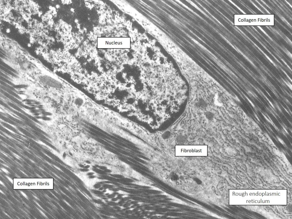
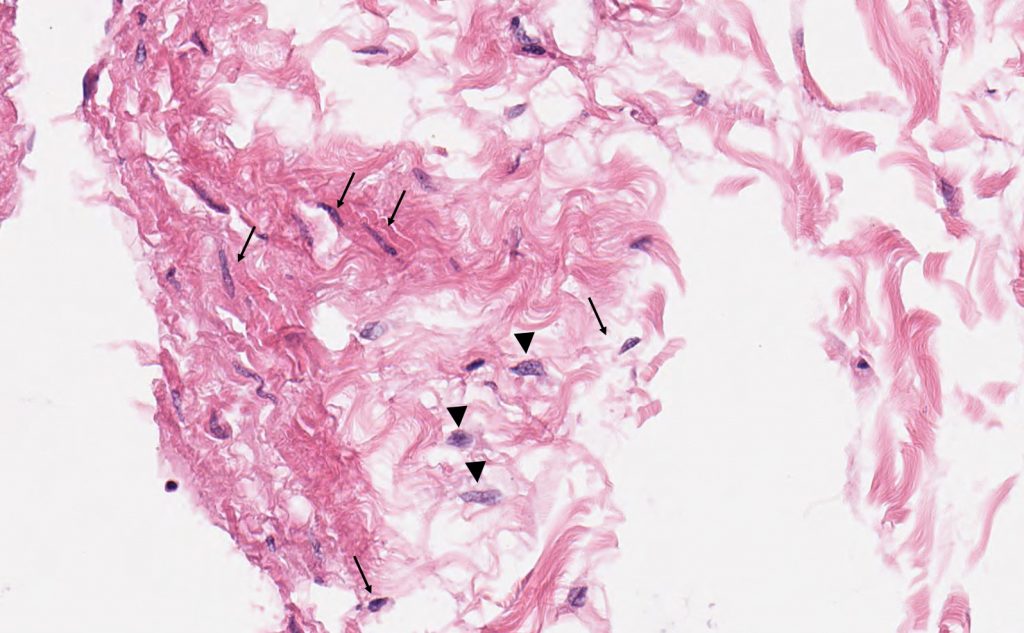
Fibroblasts
Fibroblasts are by far the most common native cell type of connective tissue. The fibroblast synthesizes the collagen and ground substance of the extracellular matrix. These cells make a large amount of protein that they secrete to build the connective tissue layer.
Sometimes the term fibrocyte is used to distinguish a small mature cell from a larger metabolically active fibroblast; however, in general convention, the term fibroblast is more common.
Macrophage
The macrophage is the connective tissue representative of the reticuloendothelial, or mononuclear phagocyte, system. This system consists of a number of tissue-specific, mobile, phagocytic cells that descend from monocytes. These include the Kupffer cells of the liver, the alveolar macrophages of the lung, the microglia of the central nervous system. The macrophage is the connective tissue version.
Macrophages phagocytose foreign material in the connective tissue layer and also play an important role as antigen presenting cells.
Other Leukocytes
Other leukocytes are frequently present in small numbers in normal connective tissue. Lymphocytes represent an adaptive immune response in tissue. These cells typically have a small amount of cytoplasm, are round and have round nuclei. It cannot be determined from routine hematoxylin and eosin staining as to whether the cells are B lymphocytes or T lymphocytes.
Neutrophils can rarely been seen in connective tissue and are more commonly observed in inflammatory states. Neutrophils look slightly different on histology than on cytology. Unlike in a cytologic preparation, the segmentations of the nuclei are difficult to see and the nucleus frequently appear as joined hyperchromatic round structures in the cytoplasm.
Mast Cells
Mast cells are granulated cells typically found in connective tissue. These cells mediate immune responses to foreign particles. In particular, they release large amounts of histamine and enzymes in response to antigen recognition. This degranulation process is protective when foreign organisms invade the body, but is also the cause of many allergic reactions.
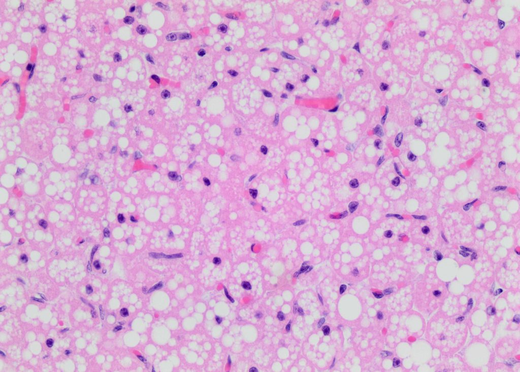
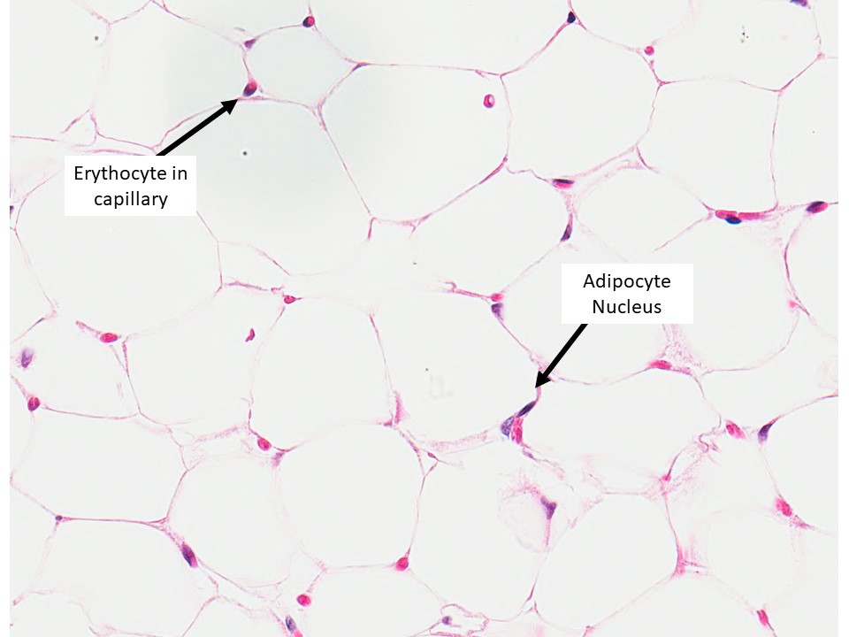
White Adipose Cells
White adipose (fat) cells are specialized for the storage of triglyceride, and occur singly or in small groups scattered throughout the loose connective tissue. When fat cells have accumulated in such abundance that they crowd out or replace cellular and fibrous elements, the accumulation is termed adipose tissue.
These cells usually contain one centrally located vacuole of lipid. The cytoplasm forms a circular ring around this vacuole and is rarely visible. The nuclei of these cells are compressed and displaced to the side. The function of white fat is to serve as an energy source and thermal insulator.
Brown Adipose Cells
Brown adipose cells are highly specialized for temperature regulation. These cells are abundant in newborns and hibernating mammals, but are typically rare in adults. Some species, such as the mouse, have abundant brown adipose stores as adults.
They have numerous, smaller lipid droplets and a large number of mitochondria, whose cytochromes impart the brown color of the tissue.