Chapter 13: Female Reproductive System
Female tubular genitalia
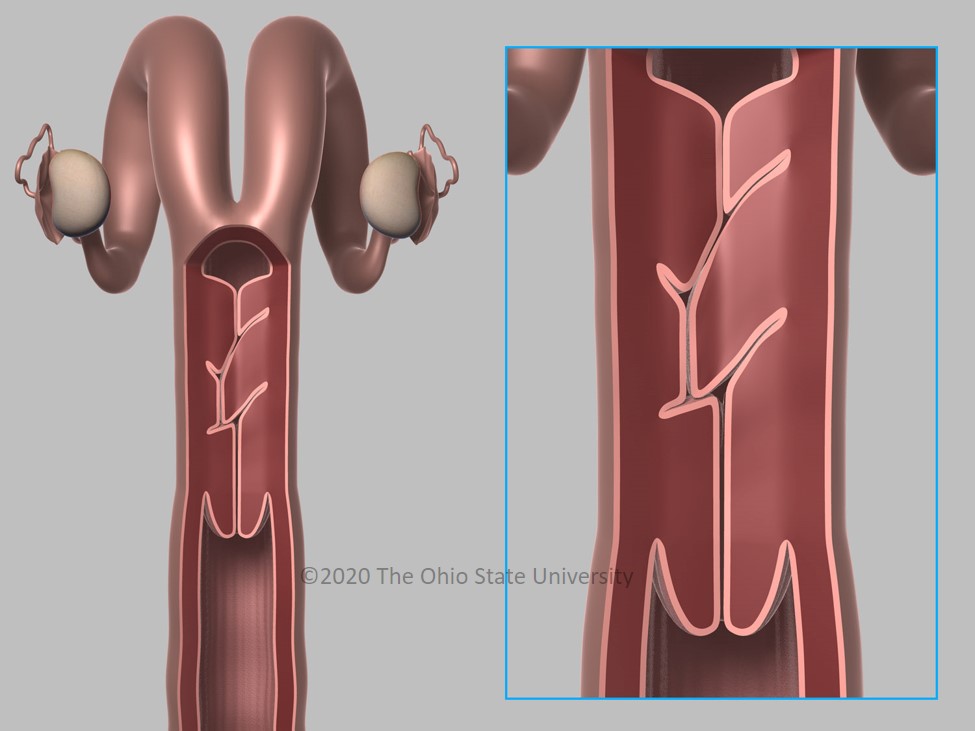
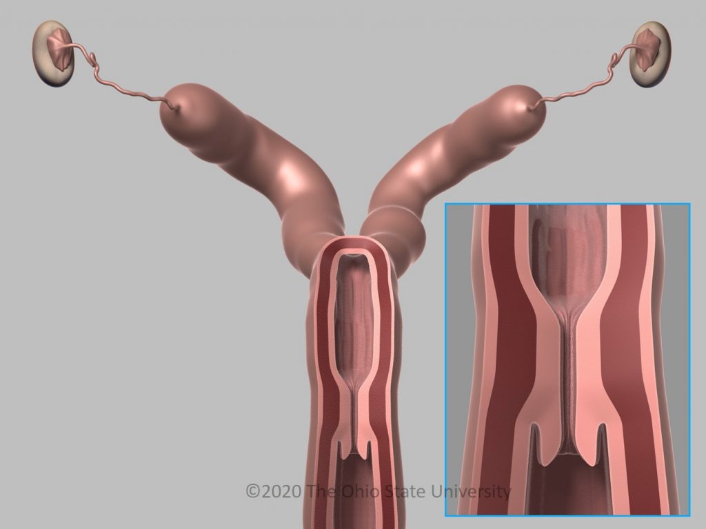
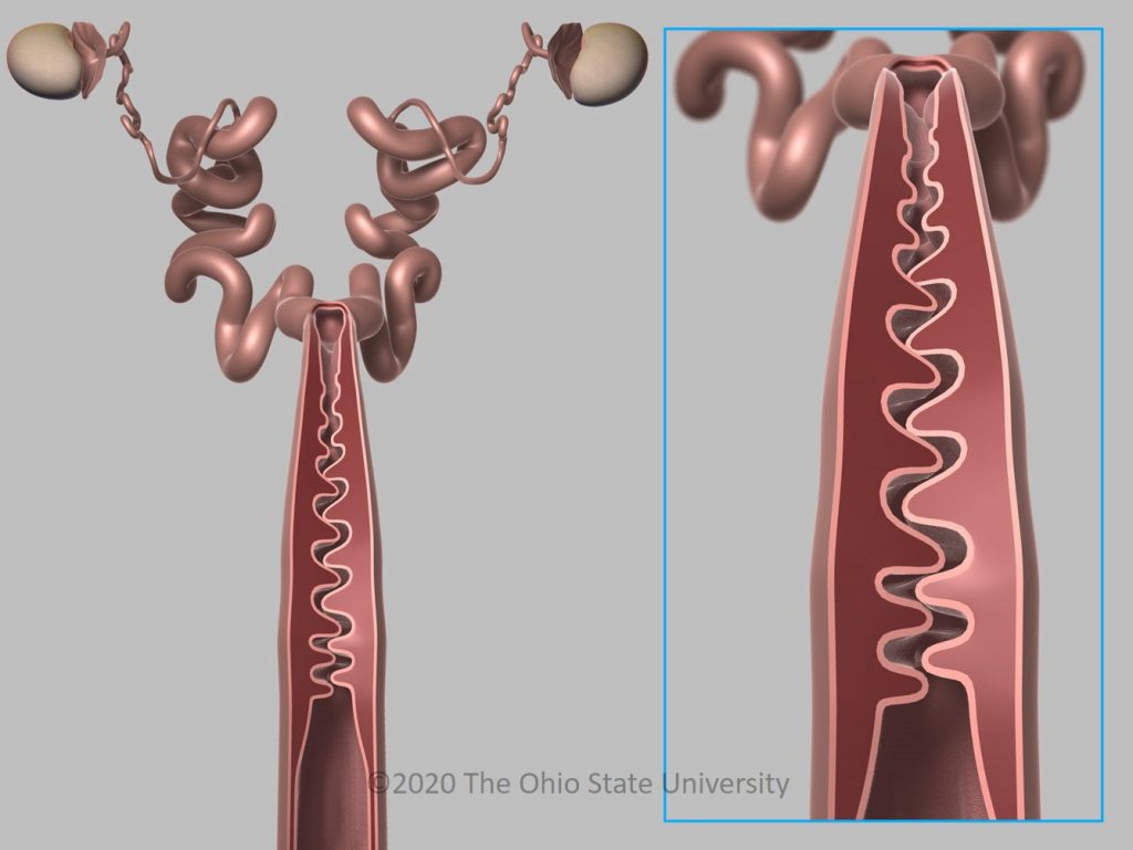
Female tubular genitalia is composed of the uterine tubes (oviducts), the uterine horns, the uterine body, the cervix and the vagina.
The uterine tubes are projections from each of the uterine horns which interface with each ovary. These structures provide transport of the oocyte towards the uterine horns and provide a site for fertilization. The uterine tubes are divided anatomically into the infundibulum, the ampulla and the isthmus. The mucosa is typically composed of ciliated columnar epithelium although in some species (bovine and porcine) it is composed of ciliated pseudostratified epithelium. The cilia move the fertilized ova towards the uterine horns.
The uterus is responsible for the maintenance of pregnancy and the transport of semen, although in some species, such as the horse, it is also the receptacle for semen. In other species, semen is deposited in the cervix or vagina. The shape of the uterus is highly species dependent. The bicornuate uterus has two large uterine horns, a uterine body and a single cervix – porcine, canine and feline, ruminants and equine. However, between species, the bicornuate uterus varies in structure. In canines, felines and swine, the uterine horns are highly developed. In cattle, sheep and horses, the uterine horns are poorly developed.
Rodents and rabbits have a duplex uterus that has two uterine openings with corresponding cervices which open into a common vagina.
The cross sectional anatomy of the uterus is divided into the endometrium, the myometrium and the perimetrium. The endometrium is typically covered with simple columnar epithelium although in some species, pseudostratified or cuboidal epithelium can be present as well. This epithelium rests on a loose collagenous tissue called the lamina propria. Beneath the mucosa, simple or branched tubular uterine glands extend from the lamina propria to the submucosa.
The endometrium is divided histologically into 3 layers.
- Stratum basalis – Deepest layer adjacent to the myometrium.
- Stratum spongiosum – Broad intermediate layer with a spongy appearance.
- Stratum compactum – Superficial layer with a compact stromal appearance.
Some species variation exists between the distribution of glands in the endometrium. The ruminant endometrium contains large vascularized protrusions (caruncles) which have relatively little glandular tissue. In addition, species variation exists in the degree of coiling at the distal ends of the glands. The depth of the gland penetration into the lamina propria and submucosa is also dependent on the stage of the estrus cycle.
The myometrium is analogous to the muscular layers found in the small and large intestine. It is composed of smooth muscle arranged in an inner circular layer and an outer longitudinal layer. Between the smooth muscle layers is a prominent vascular layer.
The perimetrium is the outermost layer and is composed of loose connective tissue, lymphatics and small arteries and veins with an overlying mesothelial surface.
FIGURE(S): Female Tubular Genitalia
The cervix lies between the uterine body and the vaginal vault where it acts as a barrier during certain phases of the estrus cycle and during pregnancy.
The mucosa of the cervix is thrown up in longitudinal folds and its mucosal composition varies between species. In the canine, the mucosa is composed of stratified squamous epithelium. The bovine cervix is contains large numbers of goblet like cells with interspersed ciliated epithelium. The structure of the cervix exhibits species variability as well. The bovine cervical mucosa contains ring-like folds which span its circumference. Longitudinal ridges are present which intersect the rings. Small ruminants (sheep and goats) have more prominent and numerous ridges which make uterine catheterization more difficult. The equine cervix is shorter (~ 6 cm) and has mucosal folds which are prominent at the caudal aspect but disappear cranially. The canine cervix is also short and has a dorsal fold of mucosa which extends into the vagina as well as transverse grooves. The porcine cervix is long (~ 25 cm) and has numerous interdigitating mucosal prominences that occlude the lumen.
The vagina is composed of four layers, the mucosa, the lamina propria, the muscularis and the adventitia. It is formed from the embryonic vaginal plate and the fused ends of the paramesonephric ducts. In the cranial vagina, the adventitia is replaced by a serosa. The mucosa in all species, except the cow, is composed of stratified squamous epithelium. The bovine vagina is lined by stratified columnar epithelium with interspersed goblet cells. Initially, the lumen of the vagina is separated from the vestibule by a thin membrane called the hymen.
The vaginal mucosa undergoes cyclical variation depending on the stage of the estrus cycle and this is of primary value in the canine. By examining the cytology from the vaginal vault, the approximate stage of estrus can be predicted.
- During anestrus (period of inactivity), the vagina contains large numbers of non-cornified, round to oval epithelial cells with large uniform nuclei. A few neutrophils may be present.
- During proestrus, large cornifying epithelial cells with shrunken or absent nuclei are present. Neutrophils are rare. Erythrocytes and bacteria may be present.
- During estrus, large cornified epithelial cells predominate. Many cells will not have nuclei. Erythrocytes are reduced.
- During diestrus, the smaller non-cornified epithelial cells reappear and neutrophils are dominant. Erythrocytes are rare.
