Chapter 10: Respiratory System
Pleura
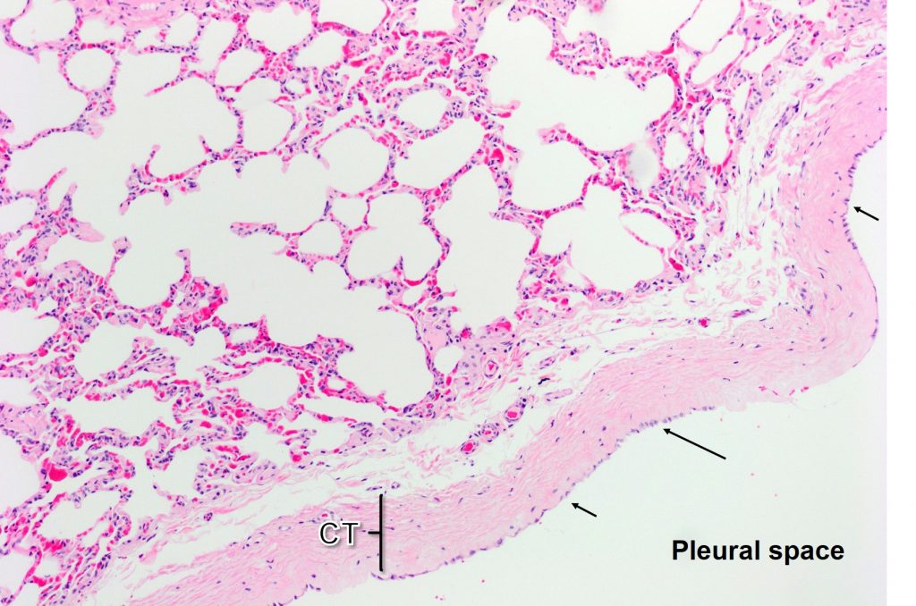
Pulmonary pleura (Visceral pleura)
The outer surface of the entire lung parenchyma is covered by the visceral pleura. The visceral pleura is composed of a thin, loose connective tissue. The outer surface is lined by specialized squamous-like cells, mesothelium. Mesothelial cells appear histologically similar to epithelial but are mesoderm derived, and express proteins consistent with both mesenchymal (stromal) cells and epithelial cells. The mesothelium produces a lubricating substance that acts to reduce friction between the visceral pleura and the pleura coating the thoracic body wall (parietal pleura) during respiration. The connective tissue of the visceral pleura is contiguous with the connective tissue of the pulmonary lobular septae that course through the pulmonary parenchyma. The visceral pleural connective tissue is typically thicker in large animal domestic species than small animal domestic species.