Chapter 9: Hepatobiliary System
Hepatic blood supply
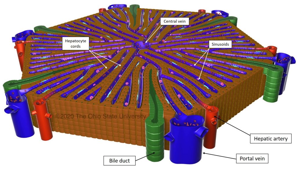
The liver is unique in that it receives blood from two sources: the hepatic artery and the portal vein. As these vessels enter the liver, their terminal branches run alongside branches of the bile ducts and course together throughout the liver parenchyma within portal triads (triad = three = hepatic artery, portal vein, bile ductule). Note that lymphatics also run within portal triads.
Portal veins are identified as larger, blood-filled spaces with a thin to inapparent vessel wall. Hepatic arteries are smaller in diameter but have a more well-developed smooth muscle wall. Blood from both the hepatic arteries and portal veins empty into the adjacent peri-portal hepatic sinusoids and flows towards the central vein.
The portal vein supplies the majority of blood to the liver. The portal circulation is a venous system that drains blood from the gastrointestinal tract, gall bladder, spleen, and pancreas. Venous blood coming from the gastrointestinal tract contains not only digested nutrients, but also toxins and, frequently, microbes. A major role of the liver in receiving this portal circulation is to metabolize these nutrients and eliminate or mitigate these toxins and microbes.
The hepatic artery delivers highly-oxygenated blood to the liver. However, once within the liver, blood from the hepatic artery and the portal vein empty into the hepatic sinusoids, combining highly oxygenated blood (hepatic arterial supply) with poorly oxygenated blood (portal vein supply). As a result, hepatocytes receive blood with reduced oxygenation. Further, when considering that blood flows from the portal triad towards the central vein, blood arriving to hepatocytes surrounding the central vein is relatively poorly oxygenated. These concepts are important in the understanding of liver pathology and how regions of the liver are differentially more susceptible to hypoxia.
The hepatic sinusoid endothelium is fenestrated, containing spaces between adjacent endothelial cells. These gaps in the endothelium allow passage of nutrients and other small compounds into the perisinusoidal space (space of Disse) where hepatocytes are able to take up these compounds. Microvilli on the surface of hepatocytes provide additional surface area for absorption of these molecules. Additionally, proteins and lipoproteins produced by hepatocytes are transferred into circulation in these perisinusoidal spaces.
