Chapter 11: Urinary system
The Glomerulus
The glomerulus is a complex web of capillaries derived from the afferent arteriole. Glomeruli can be located in the cortex of the kidney or the corticomedullary junction.
Blood that is traveling through the capillary loops is selectively filtered across the glomerular filtration barrier to form an ultrafiltrate of the plasma. This filtration barrier permits passage of small and some medium sized molecules while blocking the passage of large molecules. Filtration is driven by hydrostatic and oncotic pressure. The former pushes fluid and solutes out of the capillary lumen, whereas the latter retains fluid. The major players of the glomerular filtration barrier include: endothelium, glomerular basement membrane, and podocytes.
The glomerular endothelial cells are characterized by numerous fenestrations. Using high magnification with transmission electron microscopy (TEM), one can visualize the endothelial cytoplasm, which has numerous fenestrations or transcytoplasmic holes that allow passage of the plasma from the vasculature to the interstitium. Another important component of the endothelial cell is the glycocalyx, which covers the luminal surface of the cell. The glycocalyx is composed of negatively charged glycoproteins and proteoglycans (sugar-protein molecules). filtering out negatively charged molecules, leukocytes, red blood cells, platelets and larger proteins.
The glomerular basement membrane (GBM) is a product of the fusion of the basement membrane produced by endothelial cells and podocytes (described below). Using TEM, one can see that the GBM is trilaminar (i.e., three-layered), and these components include, from the endothelial to the podocyte aspect, the lamina rara interna, lamina densa, and lamina rara externa. This part of the filtration barrier is also negatively charged and restricts passage of larger proteins.
Podocytes are epithelial cells. Aside from providing structural and functional stability of the glomerulus, the podocyte is a vital component of the glomerular filtration barrier. They are located on the “abluminal” (i.e., opposite of capillary lumen) surface of the capillary loop. Podocytes have numerous processes, similar to tentacles, that surround the capillaries. Each process has many smaller processes (foot processes or pedicles) that are oriented perpendicularly to the capillary wall and attach to the underlying GBM. The foot processes interdigitate with foot processes of adjacent podocytes. The space between the foot processes forms the slit diaphragm. The slit diaphragm is a molecular sieve, and it provides an additional filtration barrier based mainly on size exclusion. Aside from being part of the filtration barrier, podocytes also help maintain the shape of the capillary loop shape by counteracting intraglomerular pressure. Lastly, podocytes produce vascular endothelial growth factor (VEGF) to which is a molecule that the glomerular endothelial cell needs to survive.
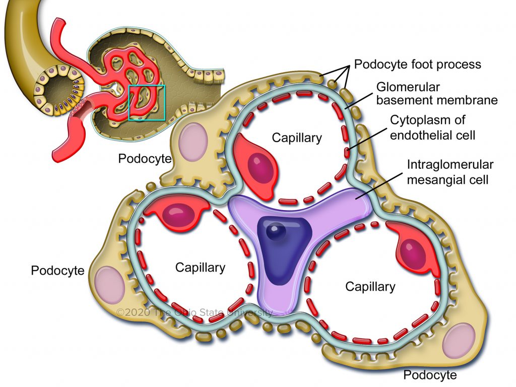
Additional structures of the glomerulus
Mesangium
Collectively, the mesangial cells and the glomerular extracellular matrix that they produce constitute the mesangium. The mesangium provides structural integrity to the glomerular tuft and modulates glomerular perfusion through smooth muscle-like activity. These cells are also involved in the production and secretion of cytokines in response to glomerular injury. Histologically, there are 2 to 3 mesangial cell nuclei beneath each capillary loop.
Bowman’s space and Bowman’s capsule
Once the ultrafiltrate is formed, the fluid enters Bowman’s space, the space between the capillary tuft and Bowman’s capsule. Bowman’s capsule is lined by parietal epithelium, which is simple squamous epithelium. The ultrafiltrate drains toward the urinary pole and enters the proximal convoluted tubule.
Efferent / Afferent Arterioles
The blood enters the glomerular tuft via the afferent arteriole and exits via the efferent arteriole. These vessels are immediately adjacent to one another at the vascular pole, which is at the opposite end of the glomerulus from the urinary pole. These vessels can contract or dilate, changing the amount of blood that flows into the glomerular tuft. Importantly, the contraction can occur in response to molecular signals from the immediately adjacent tubules.
