Chapter 15: Endocrine system
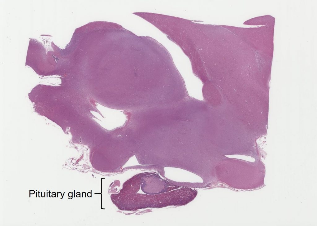
Pituitary gland
Ryan Jennings
The pituitary gland (also known as the hypophysis) is a polypoid organ that has a complex microanatomy and physical and functional interconnection with the hypothalamus (not discussed here). The physiology of the hypothalamus and its interaction with the pituitary gland is extensive, and beyond the scope of this textbook. In short, the hypothalamus is a “master regulator” of sorts, playing essential roles in body homeostasis, including temperature regulation, behavior (including thirst, hunger, and sex), inherent body rhythms, and control of the pituitary gland which, in turn, bears an immense effect on endocrine organs elsewhere. Therefore, while it is impossible to separate the functional aspects of the hypothalamus and pituitary gland, for the purpose of discussion of histology, here we will focus on the pituitary gland.
*Note: Nomenclature of the pituitary components may be confusing and we try to harmonize the language presented here, understanding that terminology may be different in other resources.
The pituitary gland is generally divided into two components with developmental and functional differences: the anterior pituitary (adenohypophysis) and the posterior pituitary (neurohypophysis). The pituitary gland is formed during fetal development via the interactions of tissue from two sources: the ectoderm of the pharynx invaginates dorsally to form the anterior pituitary gland and contacts the developing ectoderm of the brain. These developmental and functional differences in the anterior and posterior pituitary gland are apparent histologically, as well.
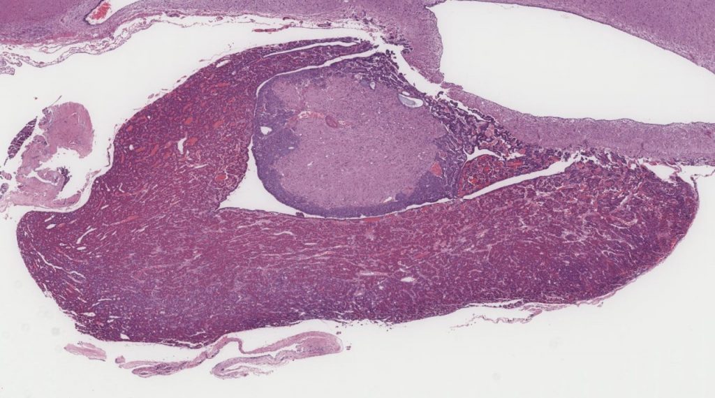
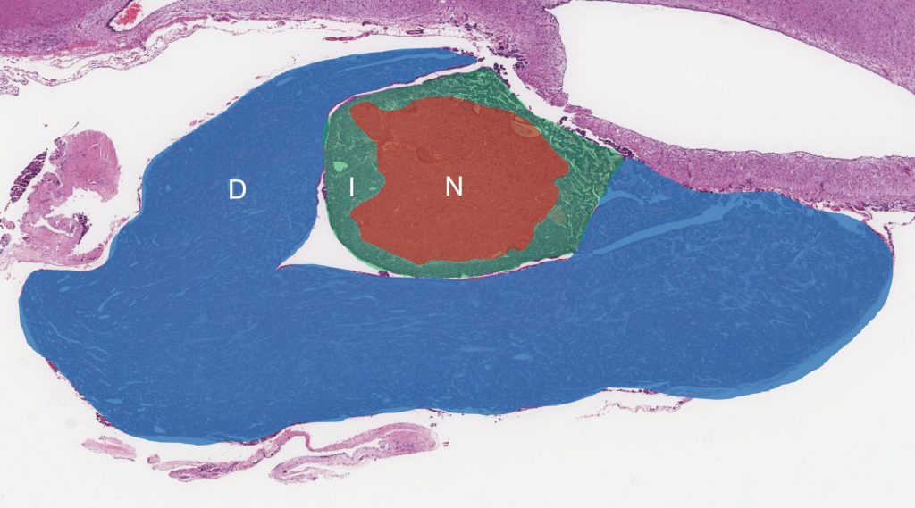
The anterior pituitary (adenohypophysis) is composed of two histologically and functionally distinct regions: the pars distalis and the pars intermedia.
The pars distalis is the largest component of the anterior pituitary gland, and is comprised of cords and trabeculae of polygonal cells arranged on a vascular stroma. Cells of the pars distalis may be categorized by their histologic appearance and by their hormone production, which do not necessarily correlate (immunostaining for hormone production is necessary). On hematoxylin and eosin staining (basic histologic stains), there are three morphologically distinct cells within the pars distalis: acidophils, basophils, and chromophobes. These are characterized by their eosinophilic cytoplasm, basophilic cytoplasm, and poorly staining cytoplasm, respectively.
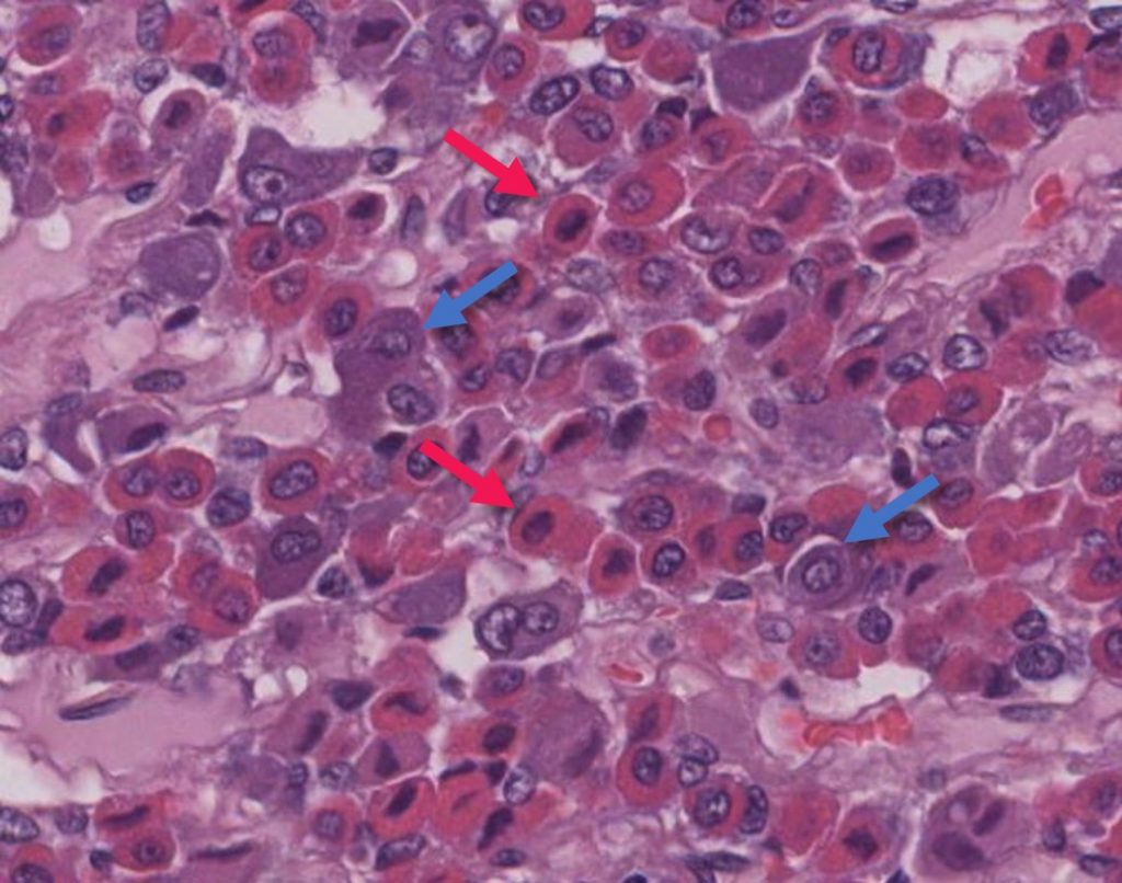
In addition to their histologic appearance, cells of the anterior pituitary may also be named according to the hormones they produce (not apparent on basic histologic stains). For example, cells that produce growth hormone are called “somatotrophs”, and cells producing ACTH are called “corticotrophs”. These can only be determined with immunohistochemical stains evaluating for specific hormone production.
The pars intermedia, the second component of the anterior pituitary, lies between the pars distalis and pars nervosa (discussed below). The pars intermedia is distinguished histologically by cords of polygonal cells with occasional formation of luminal structures lined by polygonal to cuboidal epithelial cells and containing central pink, homogeneous fluid (colloid).
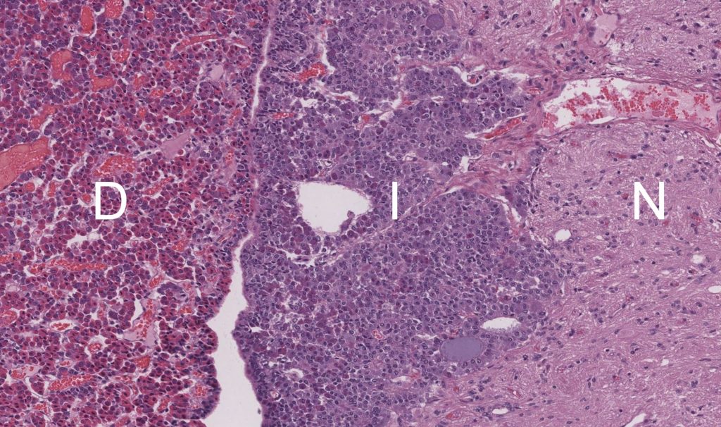
As noted above, the posterior pituitary (neurohypophysis) develops from the brain and grows ventrally to interact with and abut the anterior pituitary during development. The posterior pituitary, or pars nervosa, is histologically distinct in that it lacks the secretory polygonal cells characteristic of the anterior pituitary. Instead, the pars nervosa is composed almost entirely of non-myelinated axons, which are the elongated cellular extensions of neurons. The neuronal cell bodies themselves reside within the overlying brain (hypothalamus), and the axons extend below to form the bundle of axons that form the pars nervosa. These hypothalamic neurons are responsible for the production anti-diuretic hormone (ADH; also referred to as vasopressin) and oxytocin, which are then transported along the axons into the pars nervosa where they are secreted and enter the bloodstream.
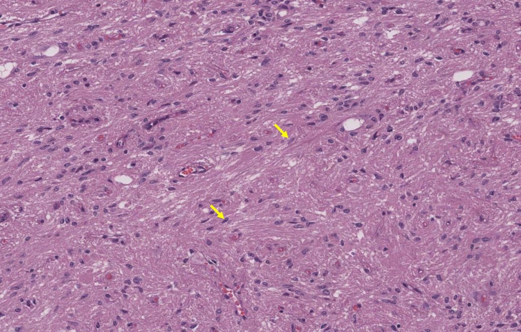
Although the pars nervosa is composed predominantly of anuclear (without nuclei) axons and small capillaries, scattered nuclei are apparent within the neuropil; these are pituicytes. Pituicytes are specialized supporting cells (glial cells) of the pars nervosa that surround the axons and facilitate secretion of ADH and oxytocin.
