Chapter 10: Respiratory System
Air conduction: Nasal cavity, Paranasal sinuses, and Vomeronasal Organ
Nasal Cavity
The paired external apertures of the nasal cavities in our domestic species are termed the external nares (nostrils). During inspiration, air enters through the nares into the most rostral segment of the nasal cavity: the nasal vestibule. The vestibule of most species is lined by keratinized stratified squamous epithelium that may contain hair follicles and other adnexa.
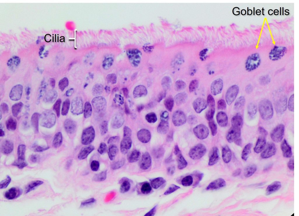
The stratified squamous epithelium of the vestibule abruptly transitions into to the characteristic respiratory epithelium. Traditional respiratory epithelium is ciliated, pseudostratified columnar epithelium. Respiratory epithelium is primarily composed of these ciliated columnar epithelial cells with basal nuclei and interspersed with moderate numbers of goblet cells. Goblet cells secrete mucus, and are characterized by cytoplasm filled with poorly staining, basophilic material (mucin).
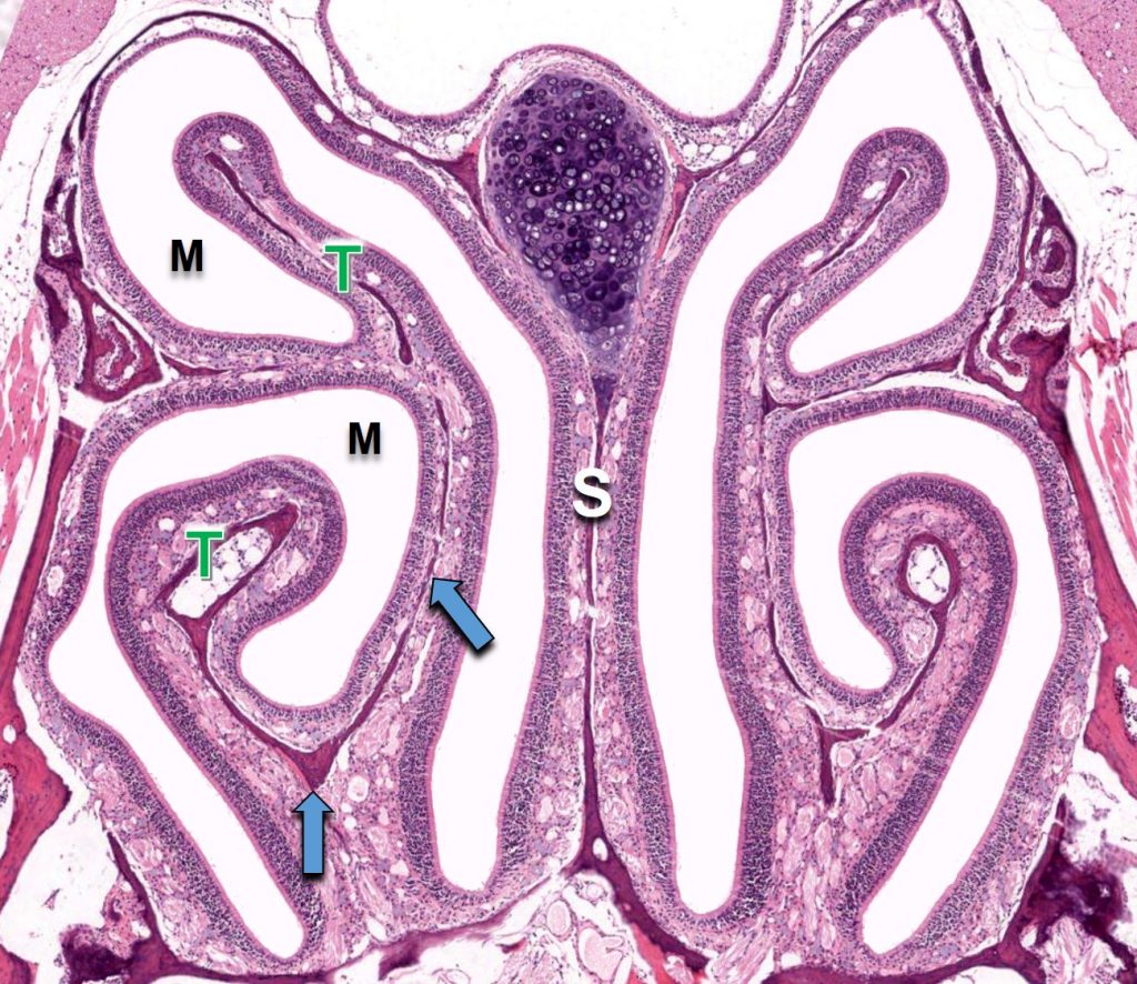
Respiratory epithelium lines the luminal surface of the nasal cavity, including the nasal turbinates. The nasal turbinates are scroll-like structures within the nasal cavity that are formed by a thin core of bone. The lamina propria and submucosa lie below the respiratory epithelium. The lamina propria may contain variable numbers of tubuloalveolar secretory glands. Similar to the salivary glands of the gastrointestinal tract, these glands may be comprised of serous cells, mucous cells, or a mixture of serous and mucous cells. These glands secrete into ducts that communicate with the surface epithelium.
Although the majority of the surface area of the nasal cavity mucosa is respiratory epithelium, portions of the more caudal regions of the nasal cavity are covered by olfactory epithelium. Whereas most of the nasal cavity is directed at physical air conduction, olfactory epithelium is a specialized epithelium that facilitates the sense of smell. Olfactory epithelium is tall, pseudostratified epithelium that contains three distinct cell types: olfactory receptor cells, sustentacular cells, and basal cells.
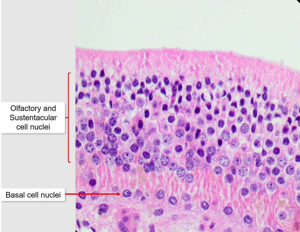
Olfactory receptor cells are modified neurons and chemoreceptor cells interspersed within the olfactory epithelium. The cell bodies of these olfactory receptor cells reside within the epithelium and have apical cilia that project onto the surface of the epithelium. These cilia are not readily distinguishable on microscopy. Nerve fibers (axons) extend from the olfactory receptor cells into the lamina propria and synapse with other axons to form collectively form olfactory nerves. These olfactory nerves, in turn, pass through perforations in the cribriform plate of the skull and enter the olfactory bulb of the brain. In this way, odors are detected by olfactory receptor cells and transmitted via axons to the brain where these odors are converted to the sense of smell.
Sustentacular cells are histologically similar to respiratory epithelium and have apical microvilli. These cells provide structural and metabolic support to the epithelium. Basal cells are cuboidal to short polygonal cells that reside adjacent to basement membrane.
Paranasal sinuses
The sinuses are bone-encased, air-filled spaces within the skull that communicate with the nasal cavity. Species such as the horse have well-developed and distinct paranasal sinuses that include the frontal, dorsal conchal, ventral conchal, rostral maxillary, caudal maxillary, and sphenopalatine. Species such as the dog have only frontal and maxillary sinuses. Regardless, the paranasal sinuses are lined by respiratory epithelium similar to that elsewhere in the nasal cavity and respiratory tract.
Vomeronasal organ
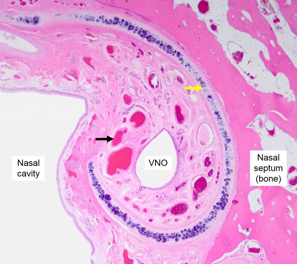
The vomeronasal organs are paired (left and right) specialized chemoreceptor organs adjacent to the nasal septum within the nasal cavity of domestic species. Structurally, the vomeronasal organ is a thick layer of sensory epithelium partially encased by cartilage and associated with an abundant vascular supply. Histologically, the sensory epithelium is similar to that of olfactory epithelium, with a thick pseudostratified epithelium that contains three populations of cells: receptor cells, sustentacular cells, and basal cells. The vomeronasal receptor cells, similar to olfactory receptor cells, are modified neurons that detect pheromones (e.g. pheromones in urine) and transmit to the brain via efferent axons. Access of air to this region of the nasal cavity is facilitated through passage through ducts in the dorsal aspect of the oral cavity (roof) of some species by the behavioral action of the flehmen response. This behavior looks different in different species. Examples include the curling of the upper lip in ungulates, chattering of the mouth in dogs (tonguing), and the open-mouthed posture of cats.
