Chapter 15: Endocrine system
Adrenal gland
Ryan Jennings
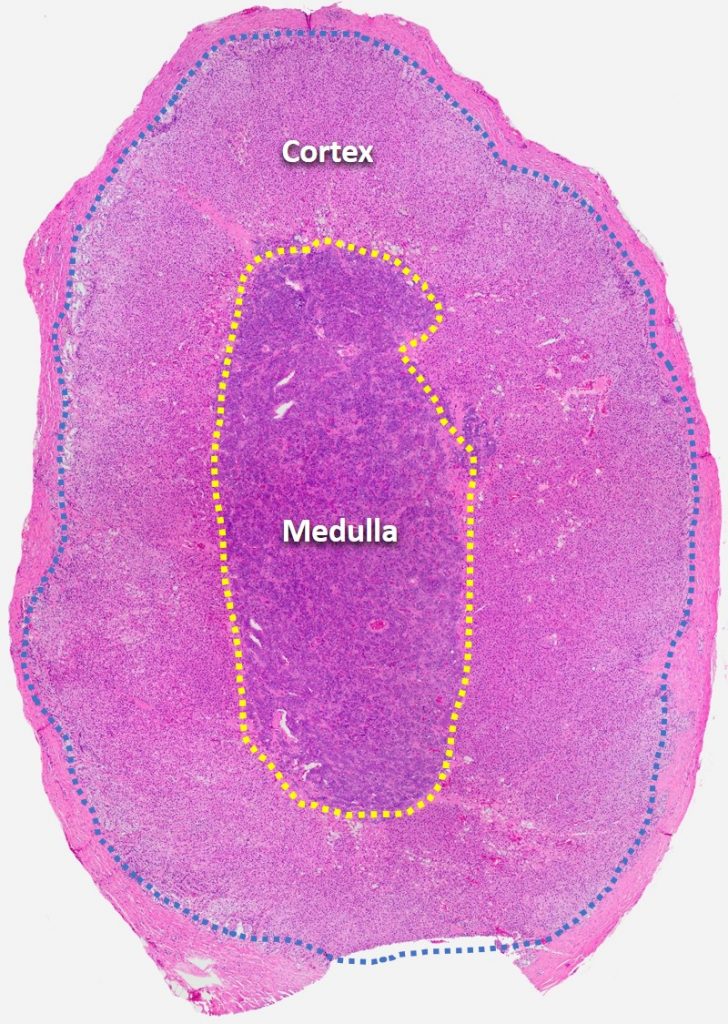
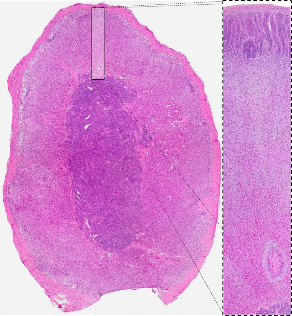
The adrenal glands of mammalian and avian species are paired organs that lie adjacent to the kidneys. The mammalian adrenal gland is surrounded by a thin fibrovascular capsule and is composed of an outer cortex that surrounds a central medulla. In avian species, cells of the adrenal cortex and medulla are intermingled, although the cytologic features of the cells are similar to that of mammalian adrenal cells.

Adrenal Cortex
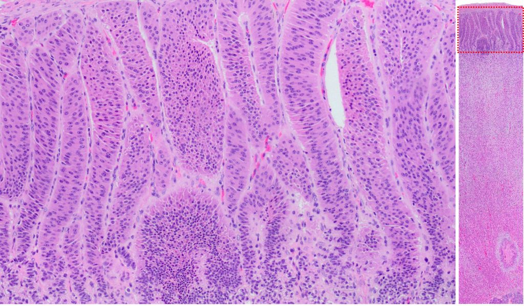
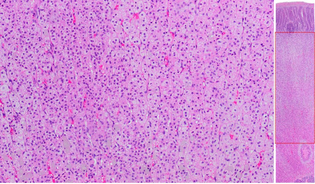
The outer adrenal cortex is composed of three functionally (and somewhat, morphologically) distinct layers, or zones, in most mammals and birds. The layers, from outermost to innermost, are: zona glomerulosa, zona fasciculata, and zona reticularis. These three layers are each responsible for producing specific steroid hormones. The epithelial cells of each layer are interspersed with an abundant network of small capillaries and scant collagenous stroma.
The zona glomerulosa, the outermost layer, is composed of trabecular arrangements of polygonal cells with moderate amounts of eosinophilic, slightly vacuolated cytoplasm, and round nuclei. Cells of the zona glomerulosa are responsible for production of mineralocorticoid hormones, primarily aldosterone, which exerts its effect on the kidneys to increase sodium retention.
The zona fasciulata is the thickest layer of the adrenal cortex, and is composed of linearly arranged trabeculae of polygonal epithelial cells with abundant eosinophilic cytoplasm is filled with small, non-staining vacuoles. The zona fasciulata primarily secretes glucocorticoid hormones, which exert their metabolic effects on numerous tissues, increasing glycogen synthesis, mobilization of lipids, an protein catabolism.
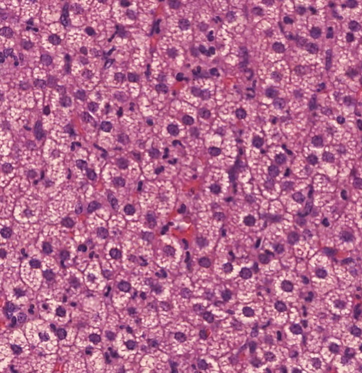
Finally, the innermost layer of the adrenal cortex, the zona reticularis, is composed of irregular cords of polygonal epithelial cells with small to moderate amounts of eosinophilic cytoplasm, with few clear cytoplasmic vacuoles. These cells are primarily responsible for the secretion of the androgen sex hormones.
Control of hormone secretion of the adrenal cortical layers is complex. The secretion of aldosterone by cells of the zona glomerulosa is controlled by the kidneys, via the renin-angiotensin system. In contrast, the zona fasciculata and zona reticularis are regulated by the pituitary gland via ACTH.
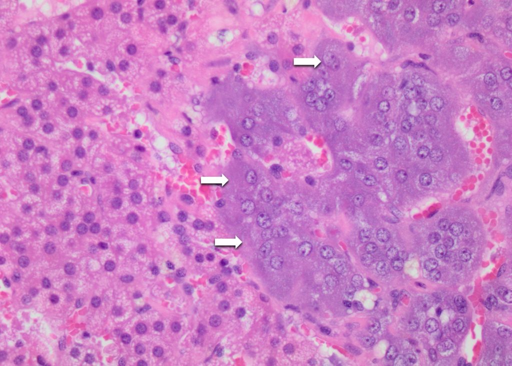
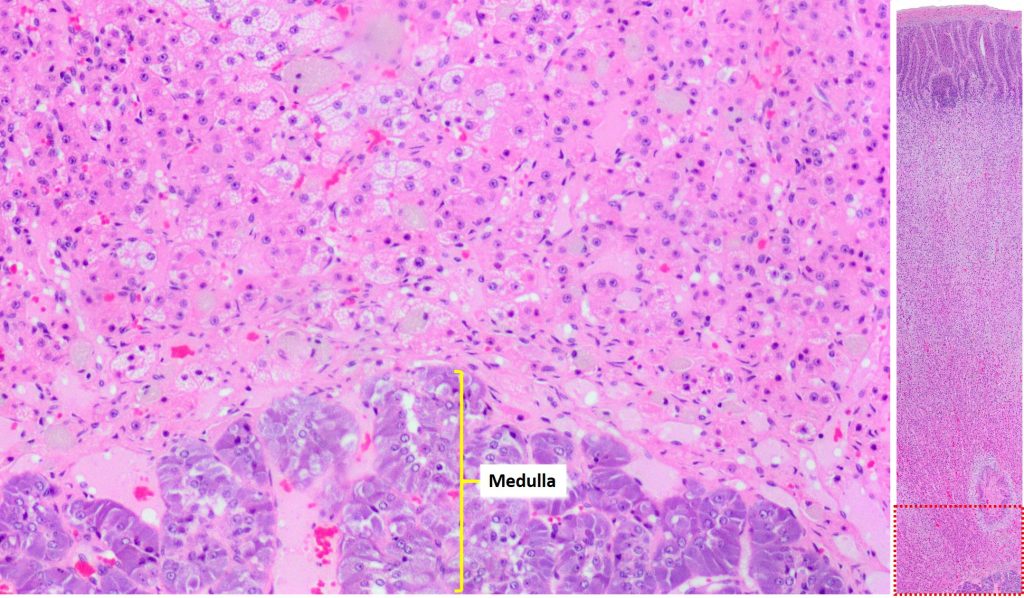
Adrenal medulla
The adrenal medulla is both histologically and functionally distinct from the adrenal cortex. The primary hormones secreted by the adrenal medulla are norepinephrine and epinephrine (also known as adrenaline). In contrast to the regulation of secretion in the adrenal cortex, secretion of these hormones is regulated by the sympathetic nervous system. The cells of the adrenal medulla are organized into clusters and irregular trabeculae of polygonal cells with moderate amounts of granular, lightly basophilic cytoplasm. These medullary cells are also referred to as “chromaffin cells”, owing to the brown staining of cytoplasmic granules when stained with chromium salts.