Chapter 11: Urinary system
Overview and Anatomy
Urine is formed in the kidney and delivered to the bladder by ureters. Urine exits the urinary bladder through the urethra. On longitudinal section, the kidney can be divided into the cortex, medulla, renal papilla and renal pelvis. The concave border of the kidney is known as the hilum (or hilus), where the renal vessels (artery and vein) and nerves enter the kidney. At this location, the renal pelvis funnels into the ureter.
In addition to dividing the kidney into cortex and medulla, it can also be separated into lobes and lobules as well as medullary rays and the cortical labyrinth. Lobes are easiest to visualize in cattle and pigs wherein every single renal papilla drains the overlying medulla and surrounding cortex. In unipapillary kidneys (e.g. carnivores), the distinction between lobes is more difficult to appreciate. A lobe can be further divided into lobules, which are composed of a single medullary ray and the surrounding cortical labyrinth, both of which are described in more detail later.
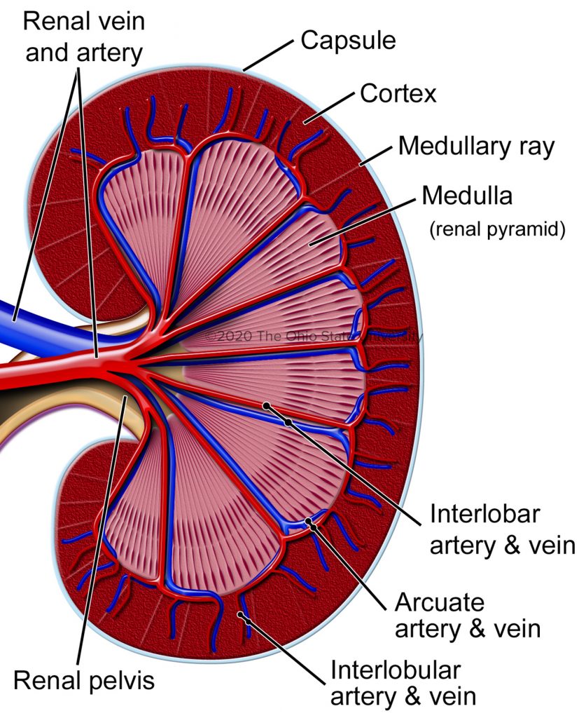
Normally, the cortex comprises approximately 2/3 of the renal parenchyma and contains most of the glomeruli and numerous cross sections of tubules. The medulla only has tubules and duct and does not contain glomeruli.
An ultrafiltrate of urine is formed in the cortex and is modified during its course through the medulla. The structures within the cortex, primarily the glomeruli, drive filtration and the tubules within the medulla are responsible for reabsorbing, excreting and concentrating the urinary filtrate.
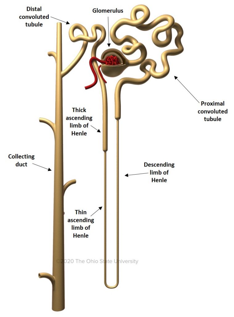
The nephron is the functional anatomic unit of the kidney. Each nephron has a glomerulus and a long tubule which becomes a duct before emptying into the pelvis. The glomeruli, drive filtration whereas the tubules are responsible for reabsorbing, excreting and concentrating the urinary filtrate. The main components of the nephron include:
- Glomerulus
- Bowman’s space (surrounded by Bowman’s capsule)
- Proximal convoluted tubule
- Proximal straight tubule
- Loop of Henle (descending limb and ascending limb)
- Distal convoluted tubule
- Collecting duct (cortical and medullary)
- Papillary duct