Chapter 9: Hepatobiliary System
Structural organization of the liver
Knowing the individual cellular and structural components of the liver, we now consider the structural organization of the liver. There are two useful ways to describe the histological architecture of the liver: lobules or acini.
Hepatic lobule (Kiernan’s lobule)
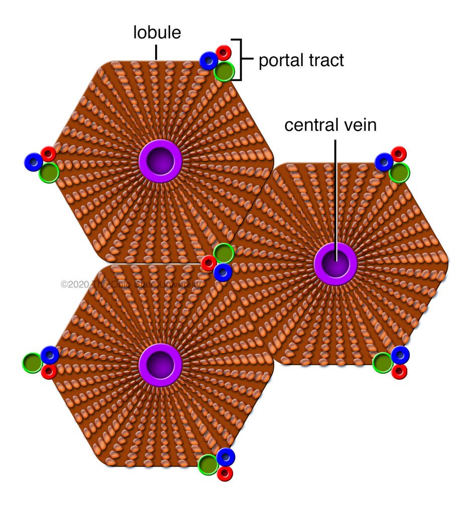
The lobular organization of the liver is one of the more histologically relevant ways to understand hepatic architecture. The two major structural landmarks to consider when reviewing the organizational aspects described below are the central veins and the portal triads. The central vein lies within the central of the hepatic lobule. Each central vein has multiple portal triads that provide blood supply to that region. Therefore, you can easily orient yourself by first identifying either a central vein (the center of a lobule) or identifying a portal tract (the periphery of a lobule).
The hepatic lobule is observed as a hexagon-like structure where the central vein is located at the center and the hepatocytes form radiating cords from the central vein, similar to the spokes of a wheel radiating from the central hub. At the outside “corners” of the hexagon are portal triads.
The hepatocellular regions within the classical lobule include portal, midzonal and centrilobular. For instance, portal hepatocytes are those closest to the portal triads, and centrilobular hepatocytes surround the central vein.
The pig liver contains increased amounts of fibrous connective tissue that bridges portal tracts, forming a band that highlights the perimeter of the lobule and illustrates the lobular organization of the liver.
Hepatic acinus (Acinus of Rappaport)
Whereas the lobular organization is based on the physical organization of structures within the liver, the hepatic acinus is based more on function. The acinus (plural: acini) highlights the hepatic blood flow and metabolic activity, and is particularly useful when considered liver disease or pathology.
The acinus is shaped as a triangle. The central vein is located at the point of the triangle and the base is at the outer edge, with the other two point of the triangle associated with portal triads. In this model, hepatocytes are defined by zones. Hepatocytes in zone 1 (also called the periportal zone) are located closest to the portal areas and are the first to receive oxygen and nutrient rich blood. These hepatocytes are metabolically active and involved in cholesterol synthesis, fatty acid oxidation and bile acid production. In contrast, hepatocytes in zone 3 (centrilobular) receive oxygen and nutrient depleted blood and are a major effector of glycolysis, lipogenesis, and xenobiotic biotransformation (highest cytochrome p450 concentration). Zone 2 (midzonal) hepatocytes have considerable regenerative potential and this region contains the greatest number of oval cells.
