Chapter 15: Endocrine system
Pancreas: Exocrine and Endocrine
Ryan Jennings
Pancreas
The pancreas is a lobulated and encapsulated gland composed of two functionally and histologically distinct components: exocrine and endocrine. In domestic species such as dogs and cats, the pancreas is a discrete organ directly adjacent to the duodenum, containing a right (proximal to the duodenum) and left limb. In large animals such as horses and cattle, the pancreas has more of a diffuse distribution within the mesentery adjacent to the duodenum.
Exocrine pancreas
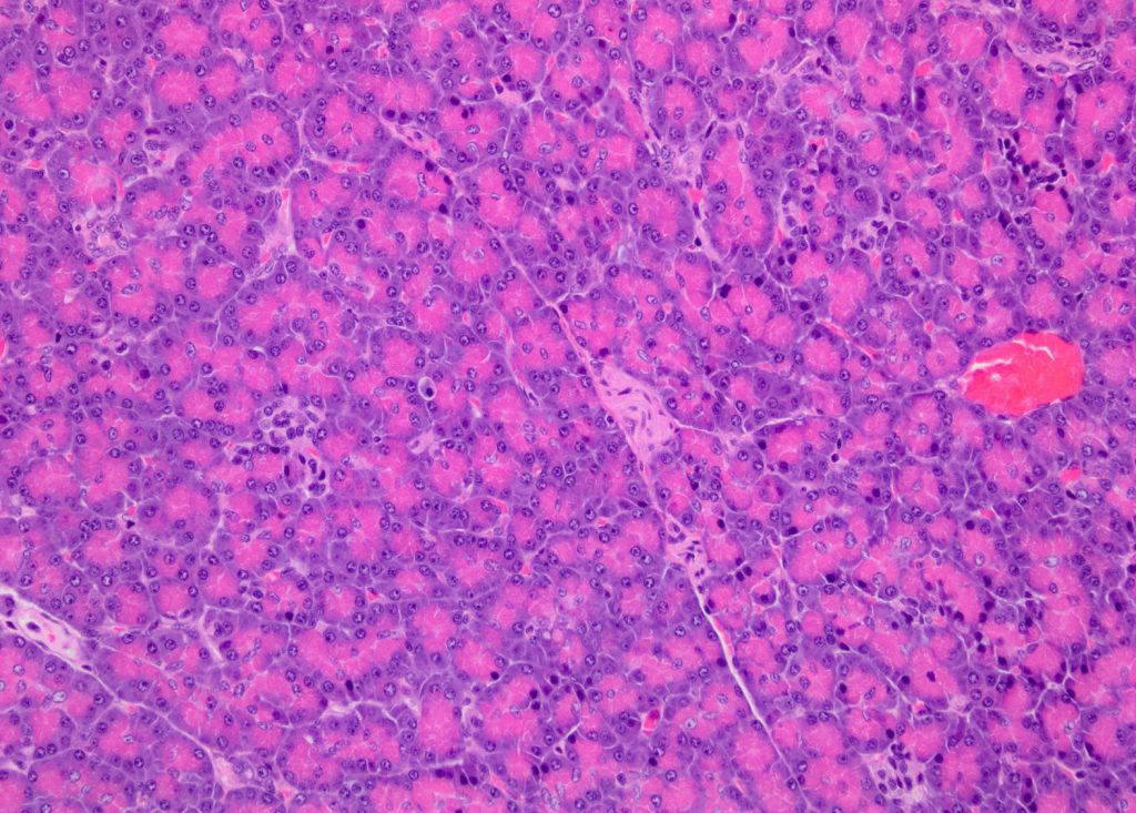
The exocrine pancreas comprises the majority cell population and volume of the pancreas. The exocrine pancreas is divided into lobules, which are separated by thin fibrovascular septa that coalesce with the pancreatic capsule which surrounds the organ. The main purpose of the exocrine pancreas is the production and secretion of diverse digestive enzymes and an alkaline fluid that neutralizes the acidic secretions of the stomach. Within the pancreatic lobules, the exocrine pancreas is composed of closely arranged acini (singular: acinus), which are luminal structures that connect and drain into a complex and branching ductular system. The pancreatic ductular system empties into the proximal duodenum via the pancreatic duct.
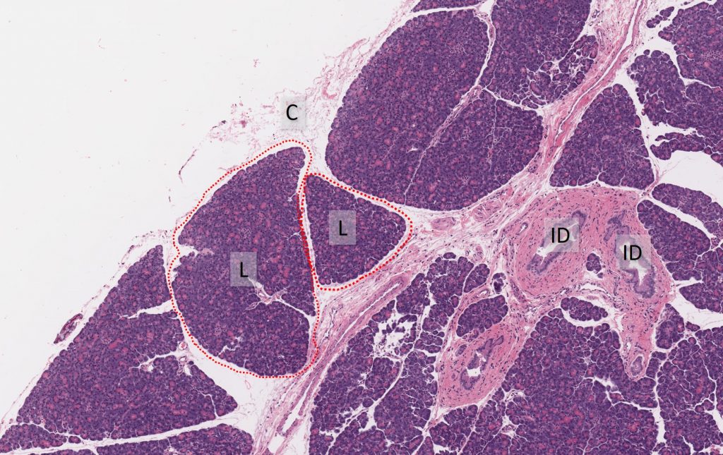

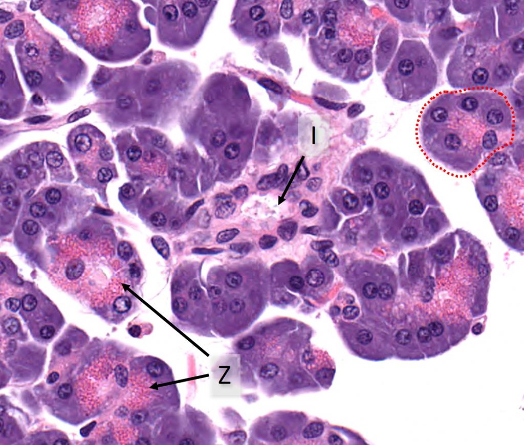
Acini are lined by polygonal to pyramidal epithelial cells (acinar cells) with basophilic cytoplmas that contains distinct, brightly eosinophilic cytoplasmic granules (zymogen granules). Zymogen granules are specialized cytoplasmic organelles that function in the sorting and secretion of pancreatic proenzymes, such as prolipase, pepsinogen and trypsinogen. These acinar cells have basally located round nuclei. The acinar cells surround small, indistinct lumina. Zymogens are secreted into the lumina which coalesce into a progressively enlarging system of ducts. The smallest ducts, intercalated ducts, are scattered between acini, and collect into larger ducts, intralobular ducts (intra- = within), which collect into even larger ducts, interlobular ducts (inter- = between). These ducts are surrounded by a supporting connective tissue stroma and lined by a single layer of cuboidal epithelial cells surrounding a central clear lumen.
Endocrine pancreas
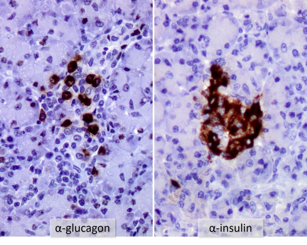
The endocrine pancreatic tissue consists of small, discrete clusters of cells, called islets of Langerhans (also called pancreatic islets), scattered throughout the exocrine pancreatic lobules. Unlike the exocrine pancreatic acini, which secrete into a ductular system, cells of the islets of Langerhans secrete directly into small fenestrated capillaries that are integrated within the islets, allowing for systemic distribution of their hormones.
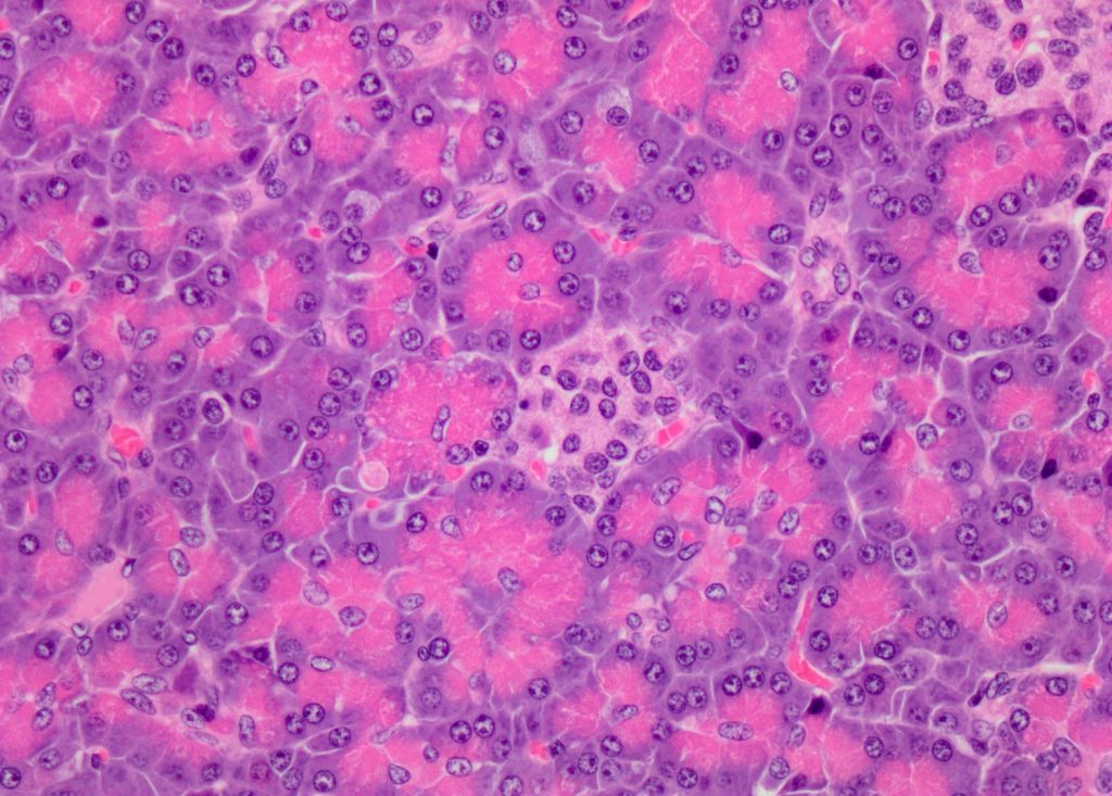
Islets of Langerhans are composed of histologically similar, though functionally distinct populations of hormone producing cells. Histologically, the cells are polygonal with moderate amounts of pale eosinophilic cytoplasm and round nuclei. Islet cells are named according to the hormones they produce. The primary hormones secreted by cells of the islets of Langerhans are insulin (β-(beta) cells) glucagon (α-(alpha) cells). Additional hormones produced and secreted include amylin, somatostatin, pancreatic polypeptide and ghrelin.