Chapter 15: Endocrine system

Thyroid gland

Ryan Jennings

The thyroid gland is a single organ present in both mammalian and non-mammalian species, but with some variation in gross anatomy.  Although a single organ, the thyroid gland of most species has distinct, anatomically separate lobes (dogs, cats, and horses).  Species such as cattle have two lobes connected by a thin strip of tissue (isthmus).  Pigs have very little lobar distinction in their thyroid gland.  Regardless of the anatomic features, the organ serves the same major functions in all species: the production of thyroid hormone and calcitonin.

The thyroid gland contains two distinct populations of hormone secreting epithelial cells: thyroid follicular epithelium, and medullary (C) cells.

The thyroid gland is surrounded by a thin fibrous capsule.  The most prominent architectural feature of the thyroid gland are the thyroid follicles.

Thyroid gland. Thyroid follicles (TF) are the predominant structures within the thyroid gland. Thyroid follicles are normally filled with a homogeneous, eosinophilic, protein-rich fluid called colloid.

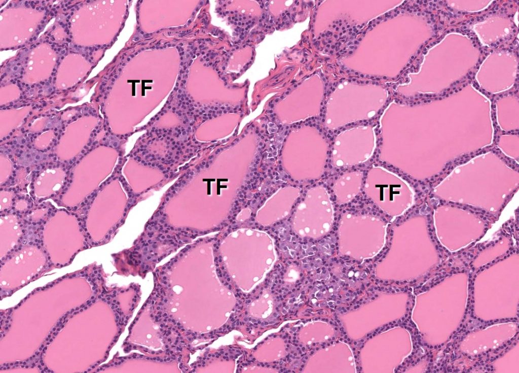
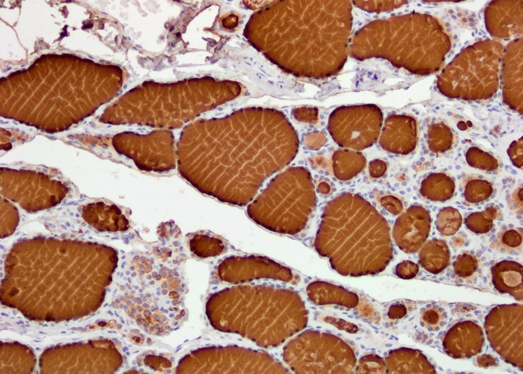
Thyroid follicles are lined by a single layer of thyroid follicular epithelium.  Thyroid follicular epithelial cells are normally cuboidal, with moderate amounts of eosinophilic cytoplasm and round, hyperchromatic nuclei.

Thyroid gland. Thyroid follicles are lined by a single layer of cuboidal thyroid follicular epithelium (arrows). The central lumina are filled with colloid (eosinophilic/pink material). Thyroid follicular epithelial cells produce and reabsorb products in colloid, which can be seen as the non-staining vacuoles, called resorption lacunae, that are associated with the luminal membrane of the epithelial cells (asterisk).

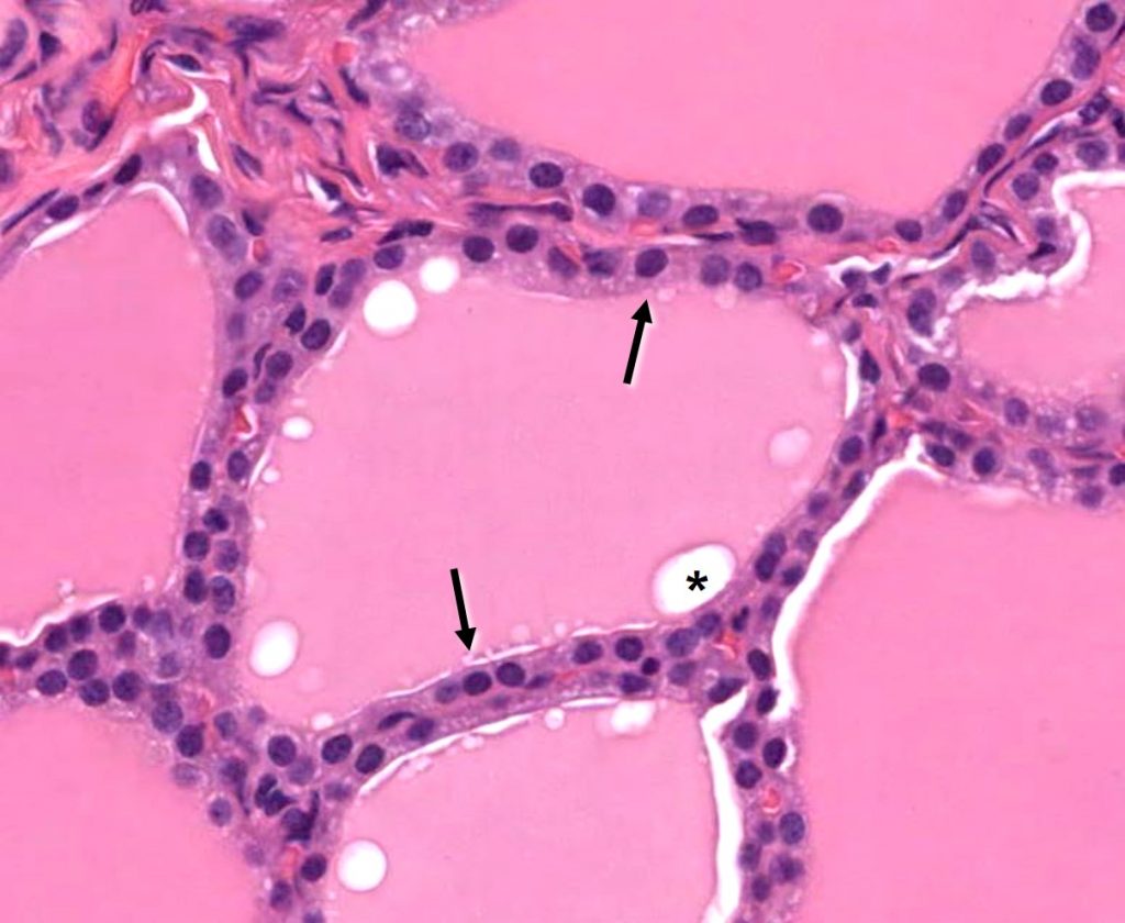
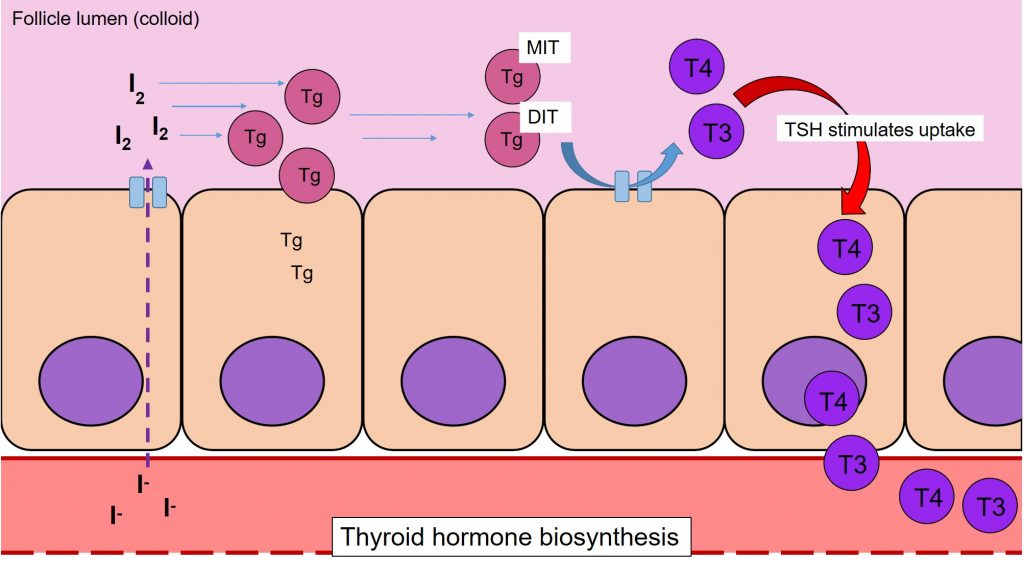
These epithelial cells surround a central lumen filled with a homogeneous, pink product called “colloid”.  Colloid is a protein rich fluid containing thyroid hormones produced by the thyroid follicular epithelium.

Thyroid gland. Immunohistochemistry for thyroid hormone. Thyroid follicular epithelial cells as well as the colloid have strong labeling for thyroid hormone, indicating a high concentration of this protein.

The thyroid follicle lumina, which contain the colloid, essentially act as a storage vat for thyroid hormone, which may then be transported across the follicular epithelium and released into capillaries adjacent to the epithelium.  This synthesis, uptake and release of thyroid hormone is mediated by TSH secreted by the pituitary gland.

Thyroid epithelium is responsible for the production of thyroid hormone. The metabolism and formation of thyroid hormone occurs on the luminal side of the thyroid follicles. Thyroglobulin (Tg) is a protein produced by the follicular epithelium and secreted into the follicular lumina. Iodine is transferred across the epithelium into the lumen where it is conjugated to thyroglobulin. Both mono- and di-iodinated forms are produced, which are then combined to produce both thyroxine (T4) and triiodothyronine (T3) (thyroid hormones). These thyroid hormones remain in the follicle lumina until thyroid stimulating hormone (TSH; produced by the pituitary gland) induces uptake and secretion of T3 and T4 into the bloodstream.


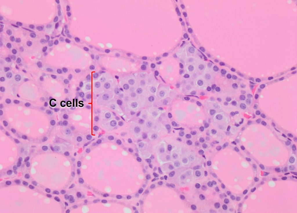
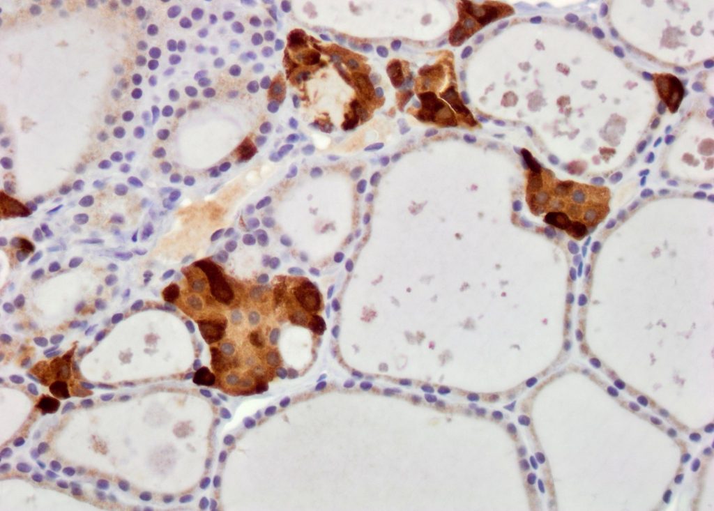
Thyroid medullary cells (also known as C cells) are polygonal cells with moderate amounts of faintly staining cytoplasm.  C cells produce and secrete calcitonin (see below), a hormone that regulates calcium/phosphate levels within the body.  C cells lie between thyroid follicles in small clusters, frequently adjacent to capillaries.

Thyroid gland. C cells (also known as thyroid medullary cells) form small clusters interspersed between thyroid follicles.


Thyroid gland. Immunohistochemistry for calcitonin. Thyroid C cells have strong labeling for calcitonin, indicating a high concentration of this protein.

License

Icon for the Creative Commons Attribution-NonCommercial 4.0 International License

Veterinary Histology Copyright © 2017 by Ryan Jennings and Christopher Premanandan is licensed under a Creative Commons Attribution-NonCommercial 4.0 International License, except where otherwise noted.

Share This Book