Chapter 6: Cardiovascular System
Capillaries and sinusoids
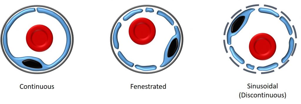
Capillaries have the smallest diameter of the vascular system. Their anatomy is such that it easily facilitates interchange of nutrients/waste between blood and organ. Capillaries are tubules less than 10 µm wide (ie. not much larger than a red blood cell). Capillaries are often collapsed in routine histologic sections and it can be difficult to appreciate this extensive, anastomosing network of small caliber vessels. Structurally, capillaries are essentially formed of endothelium supported by a basal lamina. The latter is selectively porous to small molecules approximately <70,000 MW. Loosely distributed around capillaries are pericytes that are directly apposed to the basal lamina and are capable of transdifferentiating into different cell types (ie. fibroblasts, smooth muscle cells). The most ubiquitous microcirculatory vessel in the body is the continuous capillary, with endothelial cells linked by tight junctions and intercellular gaps of approximately 1 nm. Examples of continuous capillaries are found in the lungs and brain.
Capillary structure can be yet further modified to reflect the functional needs of an organ. Fenestrated capillaries contain small cytoplasmic gaps and lie on an uninterrupted basal lamina. These vessels are more permeable than continuous capillaries, allowing easy transfer between the blood and interstitial fluid. Fenestrated capillaries are common in endocrine organs and the intestine. Porous capillaries are demonstrated in the glomerulus and allow formation of the ultrafiltrate that ultimately becomes urine.
Sinusoids are slightly larger than capillaries and are found in the adrenal glands, liver, spleen, and bone marrow. Sinusoids have large gaps between endothelial cells; the basal lamina is either discontinuous or absent. As a result, sinusoids are considerably more permeable and are capable of considerable rapid nutrient exchange. Endothelial cells lining hepatic sinusoids are interspersed by macrophages, which have an important phagocytic function.