Chapter 1: The Cell
Cell Inclusions
Cell inclusions are considered various nutrients or pigments that can be found within the cell, but do not have activity like other organelles. Examples of cell inclusions are glycogen, lipids, and pigments such as melanin, lipofuscin, and hemosiderin.
Glycogen
Glycogen is the long-term storage unit of glucose within the cell, typically in liver and muscles. Glucose molecules are connected by α(1-4) linkages and branched off by α(1-6) linkages to form the complex structure of glycogen, which also aids in rapid breakdown. Glycogen can be visualized in tissue using a periodic acid-Schiff (PAS) stain. Under an electron microscopic, two forms of glycogen exist. The first is a single spherical 15-30 nm particle (β–particle) that stain densely with lead. The second are aggregates of small particles (β-particles) called rosettes (α-particle).
Lipids
Lipid spheres in tissues are caused by an accumulation of triglycerides and appear as perfectly spherical structures. Due to processing, lipids cannot be visualized in paraffin-embedded tissues; it is assumed lipid was present when clear, spherical structures are present. On frozen tissue, lipids can be visualized using stains such as Sudan Black or Oil Red O. The material is prominent in adipocytes (fat cells), and may be found in any cell. The quantity varies with metabolic state.
Pigments
Numerous pigments can be observed in tissues and cells. Melanin is a brown pigment in hair and skin and is contained in melanosomes. These are dense ellipsoidal granules (about 0.3 x 0.7 μm) produced by specialized cells called melanocytes and are transferred to hair and skin cells to produce a pigmentary change.
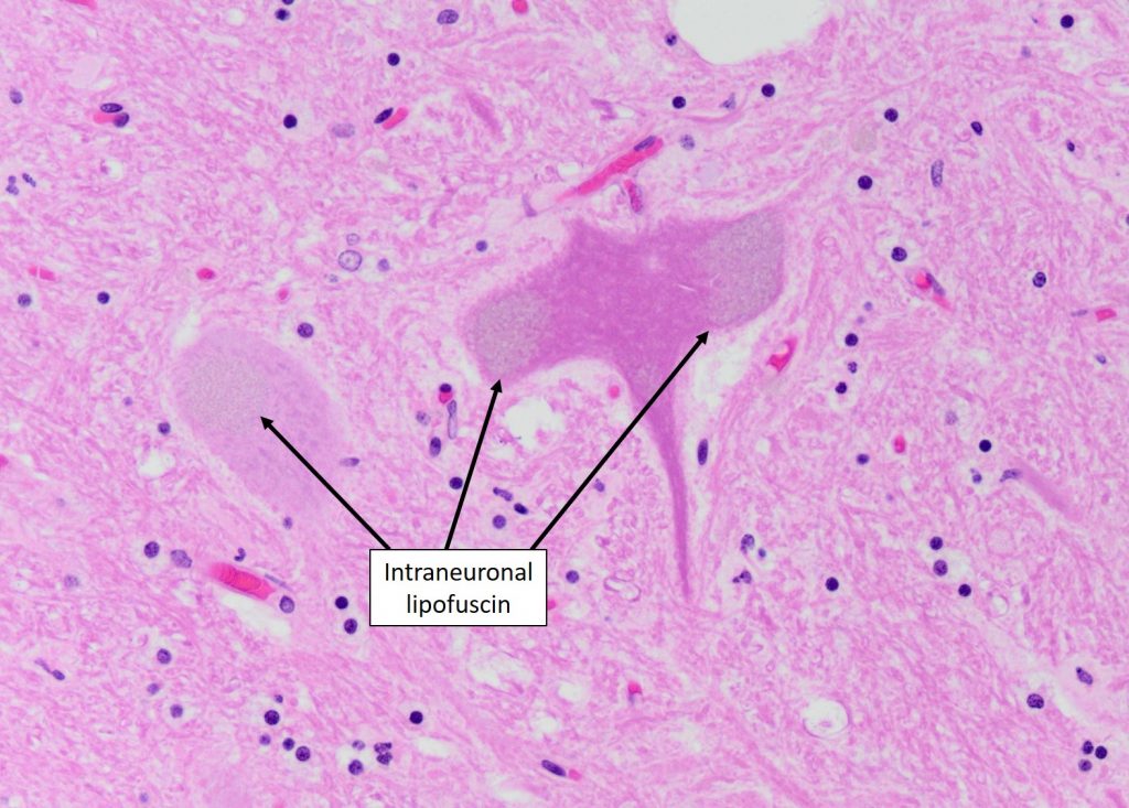
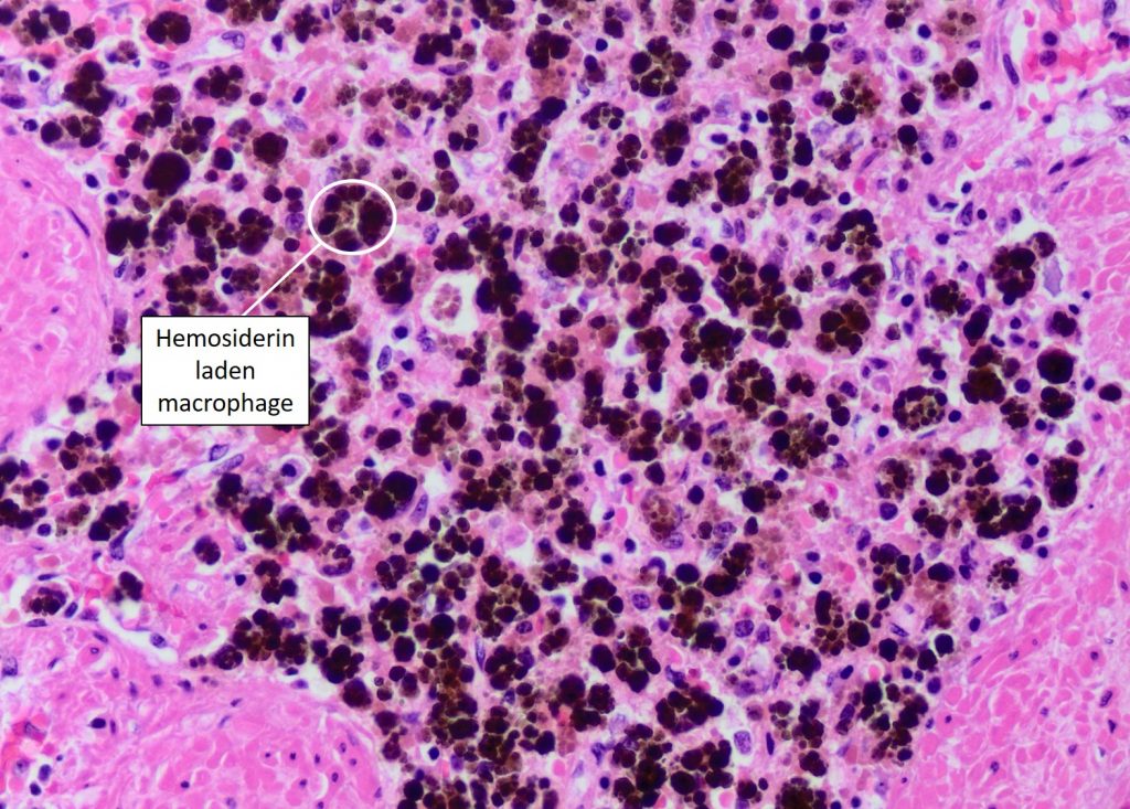
A second pigment often found in cells and tissues is lipofuscin. Lipofuscin often appears as a brown-yellow color that can be autofluorescent and accumulates over time, giving it the name “age pigment”. It is mainly found in the lysosomes of postmitotic cells. Accumulation of lipofuscin can have detrimental effects on the cell by disturbing cellular processes and leading to a degradation of cellular activity. A third pigment is hemosiderin, which is a brown pigment that contains iron. When hemoglobin breaks down, the products are phagocytized by macrophages and accumulate within the cell. Excessive hemosiderin accumulation may indicate an increase in hemolysis of red blood cells.