Chapter 8: Gastrointestinal System
General histologic anatomy of the tubular digestive tract
Distal, or aboral, to the oral cavity is the tubular digestive tract. The general histologic organization of the tubular digestive tract, from esophagus to rectum, is quite similar. These organs have a central luminal space through which digesta passes.
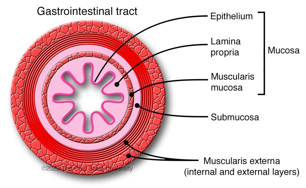
The wall of these tubular organs are organized into distinct anatomic layers; from innermost (luminal) to outermost they are:
- Mucosa
- Submucosa
- Tunica muscularis, and
- Adventitia (serosa)
The mucosa includes both the lining epithelium as well as the lamina propria and muscularis mucosa. The collagen-rich lamina propria lies directly below the mucosal epithelium (the lamina propria is similar to the dermis of skin). The mucosa also contains a thin layer of smooth muscle, the muscularis mucosa, that separates the mucosa from the submucosa. The submucosa is composed of dense collagenous stroma and contains large blood vessels, lymphatics, and nerves. The tunica muscularis is composed of two distinct layers of smooth muscle: an inner circular layer and an outer longitudinal layer. Finally, the serosa is a thin layer of fibrovascular tissue that is, externally, lined by a thin layer of flattened, specialized epithelium called mesothelial cells.
The autonomic innervation of the gastrointestinal tract functions through two distinct regions containing autonomic ganglia: the submucosal (Meissener’s) plexus, and the myenteric (Auerbach’s) plexus. This autonomic innervation enables the waves of contraction that propel food through the tubular digestive tract, also known as peristalsis. These plexi are identified as discrete clusters of neurons and nerve fibers. The submucosal plexus is located in the submucosa, whereas the myenteric plexus is located between the inner circular and outer longitudinal smooth muscle layers of the tunica muscularis.
