Chapter 2: Epithelium
Classification by number of layers of cells
Simple epithelium
Simple epithelium is a single layer of cells with every cell in direct contact with the basement membrane. It can serve many functions, including absorption, filtration and protection. The thinness of the epithelial barrier facilitates easy movement of molecules across into another compartment.
In general, simple epithelial tissues are classified by the shape of their cells. The four major classes of simple epithelium are as follows:
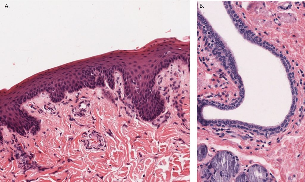
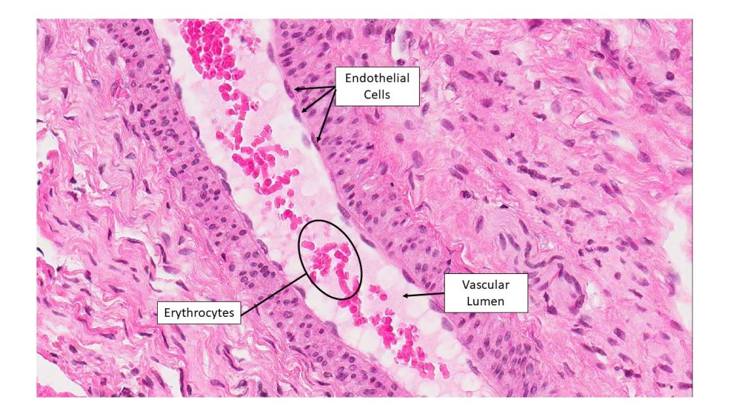
- Simple squamous: this type is found in lining areas where passive diffusion of gases occur, such as walls of capillaries and alveoli of the lungs. Endothelium is the special term used for simple squamous epithelium that lines the inside surface of a vascular structure.
- Simple cuboidal: these cells may have secretory, absorptive, or excretory functions. This type of epithelium can be seen in the tubules of kidney, and the secretory ducts of the pancreas and salivary gland.
- Simple columnar: found in areas with extremely high secretory activity (as in the inside lining of the stomach), or absorptive activity (as in small intestine). This type of epithelium often possess apical modifications such as the microvilli in the small intestine, or cilia found in some areas of the female reproductive tract).
- Pseudostratified epithelium: this type of epithelium is almost exclusively located in the larger respiratory airways of the nasal cavity, trachea and bronchi. For this reason, it is often also called respiratory epithelium.
FIGURE(S): Epithelium Classification by Number of Layers of Cells
There are two notable examples of simple squamous epithelium that have unique names. The first, endothelium, refers to the simple squamous epithelium that lines in the inside surface of all vascular structures. Endothelium is a critical component of vascular structures that will be discussed in the cardiovascular section. The second, mesothelium, refers to the simple squamous epithelium that lines the outside surface of many organs in the abdominal and thoracic cavities. It forms a component of the serosa, the lining of abdominal and thoracic organs as well as the parietal peritoneum and pleura.
Stratified epithelium
Stratified epithelium differs from simple epithelium in that it is multilayered. It is therefore found where body linings have to withstand mechanical or chemical insult such that layers can be abraded and lost without exposing subepithelial layers.