Chapter 4: Muscle
Skeletal Muscle
Function
One of the most important functions of skeletal muscle is to contract and cause movement. Skeletal muscles act not only to produce movement but also to stop movement. Constant small contractions of the skeletal muscle are needed to hold a body upright in any position, even at rest. Muscles also prevent excess movement of the bones and joints, maintaining skeletal stability and preventing skeletal structure damage or deformation. Skeletal muscle is located throughout the body at the openings of internal tracts to control the movement of various substances. This muscle allow voluntary control of specific functions, such as swallowing, urination, and defecation. Skeletal muscles also protect internal organs (particularly the abdominal organs) by acting as an external barrier or shield to external trauma and by supporting the weight of the organs.
Skeletal muscles contribute to the generation of heat, primarily by shivering or intense bursts of movement. Muscle contraction requires energy, and when ATP is broken down, heat is produced. This heat is very noticeable during and following intense active movement such as running. In this case, sustained muscle movement causes body temperature to rise. Similarly, when environmental temperatures are low, random skeletal muscle around the appendicular skeletal produces random skeletal muscle contractions to generate heat (shivering).
Structure
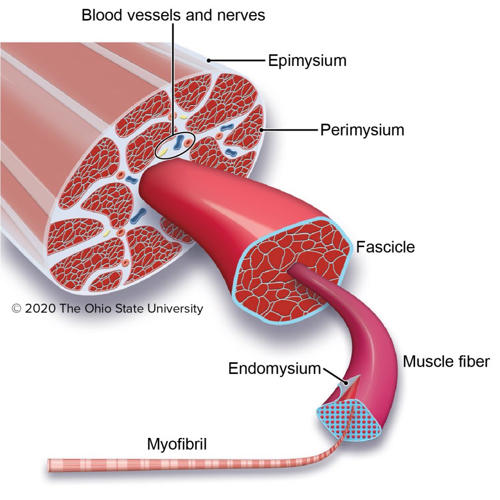
Each skeletal muscle is an organ that consists of various integrated tissues. These tissues include the skeletal muscle fibers, blood vessels, nerve fibers, and connective tissue. Each skeletal muscle has three levels of connective tissue that enclose it. These levels of connective tissue provide structure to the muscle as a whole, and also compartmentalize the muscle fibers within the muscle in to groups. Each large muscle is wrapped in a sheath of dense, irregular connective tissue called the epimysium, that holds muscle fascicles in a group and also allows a muscle to contract while maintaining its structural integrity. This is the outermost layer. The epimysium also allows the muscle to move independently from other muscle groups and other types of tissue. Most individual muscles that you learn in gross anatomy are surrounded by epimysium.
Muscle fiber is a term synonymous with muscle cell. The muscle fiber is a cell with multiple peripherally located nuclei that runs the ENTIRE length of the actual muscle. Inside each skeletal muscle, muscle fibers are organized into individual bundles (called fascicles), by intermediate layer of connective tissue called the perimysium. Inside each fascicle, each muscle fiber is encased in an extremely thin connective tissue layer called the endomysium.
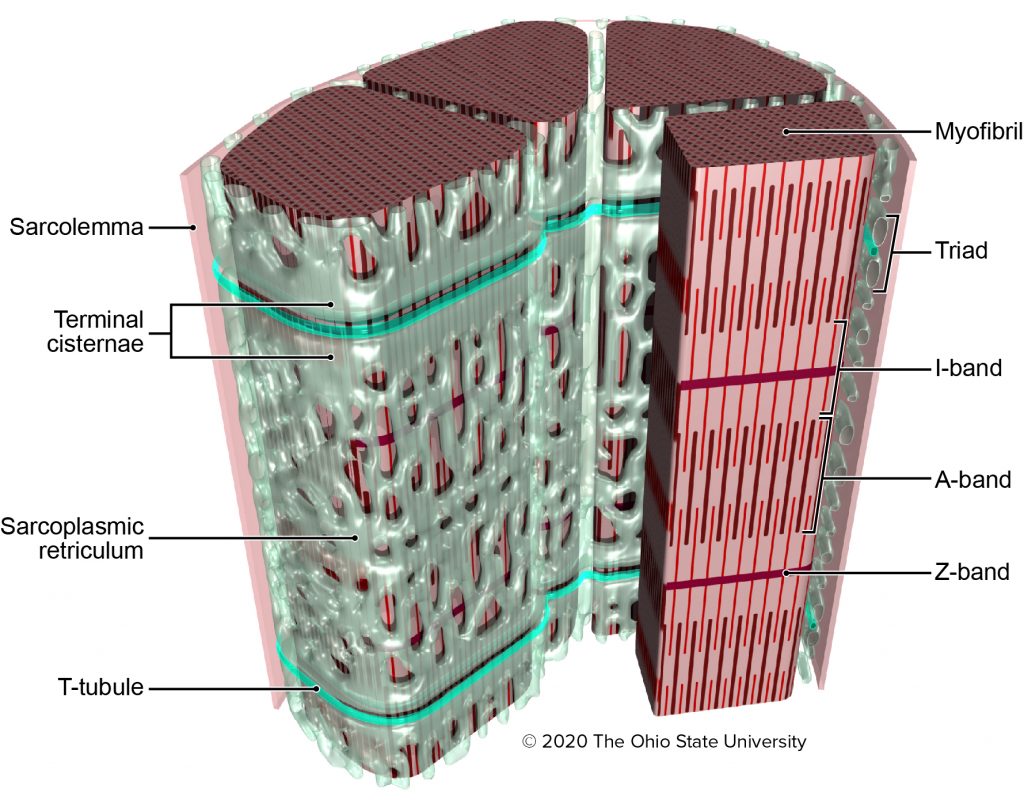
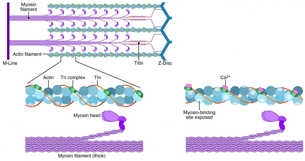
Some terminology associated with muscle fibers are rooted in the Greek sarco, which means “flesh.” The plasma membrane of muscle fibers is called the sarcolemma, the cytoplasm is referred to as sarcoplasm, and the specialized smooth endoplasmic reticulum that stores and controls the release of calcium ions (Ca++) is called the sarcoplasmic reticulum (SR). The functional unit of a skeletal muscle fiber is the sarcomere, a highly organized arrangement of the contractile myofilaments actin (thin filament) and myosin (thick filament), along with other support proteins. Individual sarcomeres are joined together in a long chain called a myofibril and attaches to the sarcolemma at its end. Myofibrils are bundled together within the sarcoplasm. In addition, there are invaginations of the sarcolemma into the sarcoplasm that surround the myofibrils. These are referred to as t-tubules. The t-tubules system conveys the action potential from the sarcolemma into the cell in order stimulate the myofibrils.
Distinct lines or bands in the sarcomere are apparent where the myosin and actin filaments overlap as well as where the sarcomere connect to each other consecutively. This is what gives skeletal muscle its striated appearance on histology. In fact, skeletal muscle and cardiac muscle (because cardiac muscle also has sarcomeres) are also known as striated muscle. As the sarcomeres and thus, the myofibrils contract, the entire muscle cell contracts. Because the actin and its troponin-tropomyosin complex (projecting from the Z-discs toward the center of the sarcomere) form strands that are thinner than the myosin, together it is called the thin filament of the sarcomere. Because the myosin strands and their multiple heads (projecting from the center of the sarcomere, toward but not all to way to, the Z-discs) have more mass and are thicker, they are called the thick filaments. As mention before, in order for a sarcomere to contract, calcium released from the sarcoplasmic reticulum after a stimulatory action potential from the T-tubules is generated. Calcium interacts with molecules on the actin filaments, troponin and tropomyosin, which normally block myosin binding sites on actin. ATP binding to myosin causes release of the myosin head from actin.
Contraction
Contraction of the sarcomere involves several steps. In order to understand these step, you must understand that myosin head has several important characteristics.
- It has ATP-binding sites into which fit molecules of ATP. ATP represents potential energy.
- It has actin-binding sites into which fit molecules of actin.
- It has a “hinge” at the point where it leaves the core of the thick myofilament. This allows the head to swing back and forth, and this motion is what causes muscle contraction.
During contraction the following steps occur:
- The activated myosin head attaches to the actin binding site; contraction refers to the activation of myosin’s cross bridges – the sites that generate the force
- The phosphate group, P, on the myosin head falls off
- The myosin head then produces a power stroke which results in the sliding of the filaments forcing the thin filaments to move towards each other. The ADP on the myosin also falls off.
- Once the power stroke is complete, ATP again attaches to the myosin head causing the head to detach from the actin site and return to its original position.
- ATP attached to the myosin head is split by ATPase causing the myosin heads to be activated again; the cycle can then be repeated over and over again as long as calcium and ATP are present. Multiple cycles of this process are often necessary for full muscle contraction.
