Chapter 8: Gastrointestinal System
Stomach – additional interspecies variations
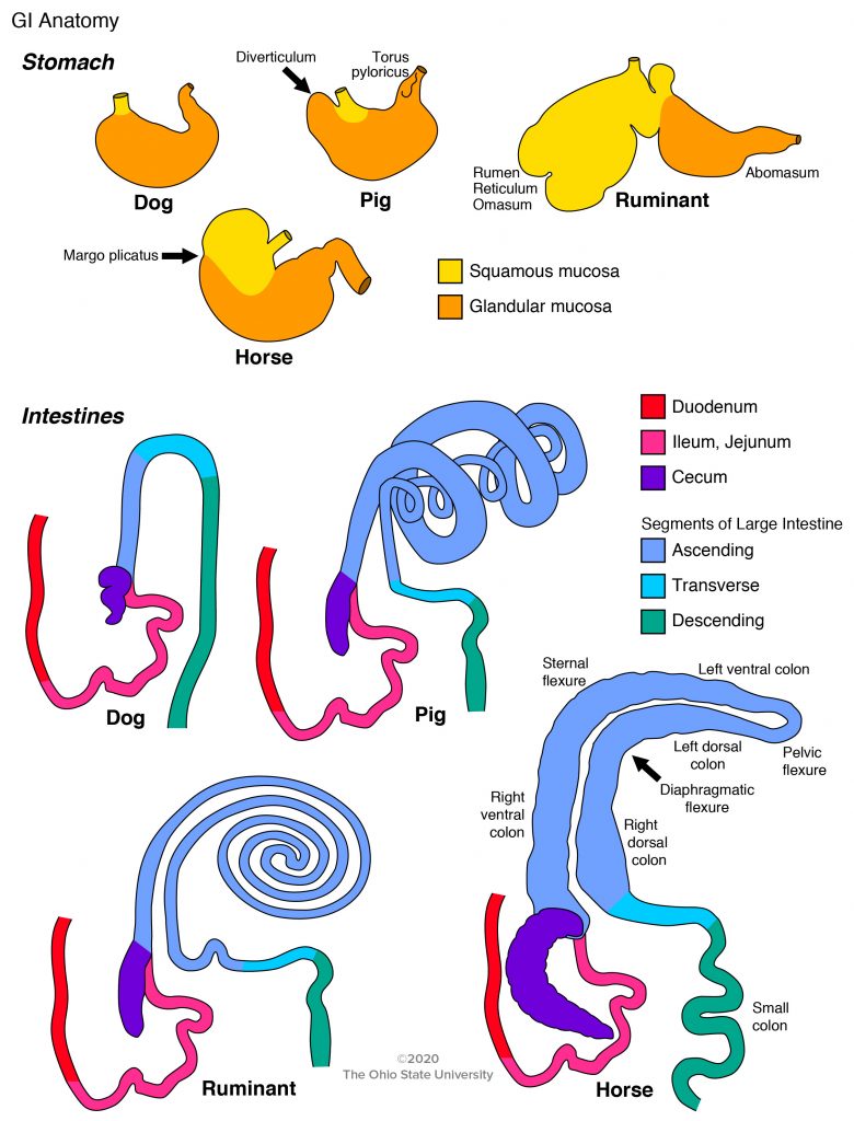
Horses
The equine stomach is composed of a proximal nonglandular mucosal region and an aboral glandular gastric mucosa. The anatomically distinct demarcation of these regions is referred to as the margo plicatus. The nonglandular mucosa is lined by stratified squamous epithelium, whereas the glandular portion is analogous to that of carnivores.
Pigs
In pigs, the gastric region immediately aboral to the esophageal sphincter is a well-delineated segment of nonglandular mucosa, the pars esophagea. The pars esophagea is lined by stratified squamous epithelium. The remaining gastric mucosa is glandular and analogous to that of carnivores.
South American Camelids (Llama, Alpaca)
Similar to the stomach of ruminants, the camelid stomach is anatomically divided into specialized compartments that serve to facilitate digestion of fibrous ingesta (foregut fermentation). The stomach of camelids is divided into three compartments: compartment 1 (C1), compartment 2 (C2), and compartment 3 (C3). Compartment 1 (C1) is analogous to the ruminant rumen, and is a large volume saccule that acts as a vat for microbial fermentation. In contrast to the rumen, C1 is lined primarily by glandular mucosa with regions of non-glandular stratified squamous epithelium. The C1 also contains numerous small out-pouchings, or saccules. The C2 is composed of both glandular mucosa and non-glandular stratified squamous epithelium. Finally, C3 is functionally and histologically analogous to the ruminant abomasum and canine stomach and is entirely glandular.
Chickens (and other fowl)
The proventriculus is glandular stomach that is functionally, but not histologically, analogous to the canine stomach. The wall of the proventriculus contains numerous large, well-circumscribed glands (proventricular glands), each formed by collections of branching tubules lined by regions of columnar mucous cells (microscopically similar to those of the gastric pits) and cuboidal to low columnar secretory cells. These secretory cells have eosinophilic granular cytoplasm and produce pepsinogen and hydrochloric acid, thereby serving the roles of both the chief and parietal cells of the canine stomach, respectively. The proventricular glands open to the luminal surface through apertures in the mucosal surface. The superficial proventricular mucosal epithelium is primarily composed of mucous cells.
The gizzard is the compartment immediately aboral to the proventriculus and is distinguished by a remarkably well-developed and thick smooth muscle wall that contributes to mechanical breakdown of ingesta. The gizzard is generally more developed in bird species with diets consisting of tough material (grains) and less so in species with more soft diets (fruit). Histologically, the mucosa is composed of vertically oriented tubular glands lined by columnar epithelium. The superficial (luminal) surface of the mucosa is lined by a thick layer of homogeneous, glassy eosinophilic to slightly basophilic material called koilin. Koilin forms a tough protective coating over the mucosal surface and contributes to the ability of the gizzard to grind particles of ingesta. The molecular composition of koilin is similar to but distinct from that of keratin. The koilin is produced by the tubular glands of the gizzard, which interdigitate with the koilin to provide structural integrity.
