Chapter 2: Epithelium
Classification by function of epithelium
Epithelium can have two functions. They can act as a lining (lining epithelia) and/or form secretory structures (glandular epithelium).
Lining Epithelia covers the free surfaces of the body (internal and external) and cavities. Examples of this include epidermis, lining of the gastrointestinal tract and ducts. Their position in contact with the environment gives them great importance in regulating the composition of the body by controlling the movement of materials in and out. Tissues that line the inside of the mouth, the esophagus and part of the rectum are composed of nonkeratinized stratified squamous epithelium. Other surfaces that separate body cavities from the outside environment are lined by simple squamous, columnar, or pseudostratified epithelial cells. Other epithelial cells line the insides of the lungs, the gastrointestinal tract, the reproductive and urinary tracts, and make up the exocrine and endocrine glands. The outer surface of the cornea is covered with fast-growing, easily regenerated epithelial cells. Endothelium (the inner lining of blood vessels, the heart, and lymphatic vessels) is a specialized form of epithelium. Another type, mesothelium, forms the walls of the pericardium, pleurae, and peritoneum.
Glandular epithelium is an invagination or aggregation of epithelium that forms a solid tissue structure. Glandular tissue is formed from glandular epithelium that forms structures from the infolding of epithelium and subsequent growth in the underlying connective tissue.
FIGURE(S): Diagram of Invaginated Epithelium to Form a Gland
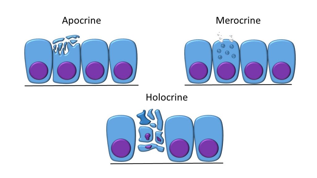
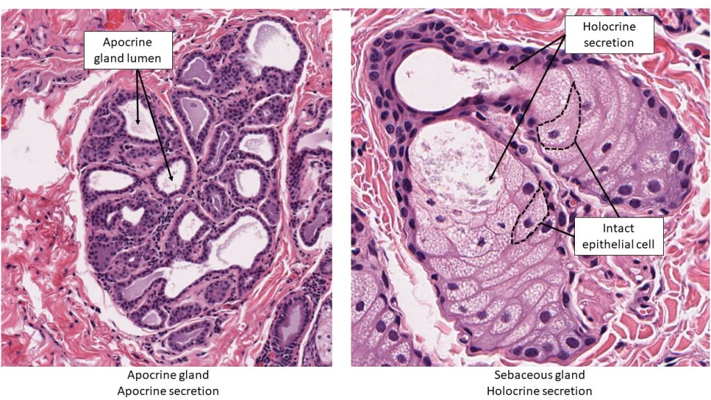
Glands can be broadly classified into two types : endocrine and exocrine. Endocrine glands secrete their product into the extracellular space where it is rapidly taken up by the blood vascular system. Exocrine glands secrete their products into a duct that then delivers the product to the lumen of an organ or onto the free surface of the epithelium. Exocrine glands are further characterized by the method in which they secrete product. Merocrine secretion involves the exocytosis of secretory vesicles to the lumen of the gland. Holocrine secretion involves the death of an entire cell within a gland which subsequently sloughs off and releases content into the lumen of the gland. Apocrine secretion involves the release of budding vesicles off the epithelial cells into the lumen of the gland.