Chapter 10: Respiratory System
Gas exchange: Alveoli
Alveoli
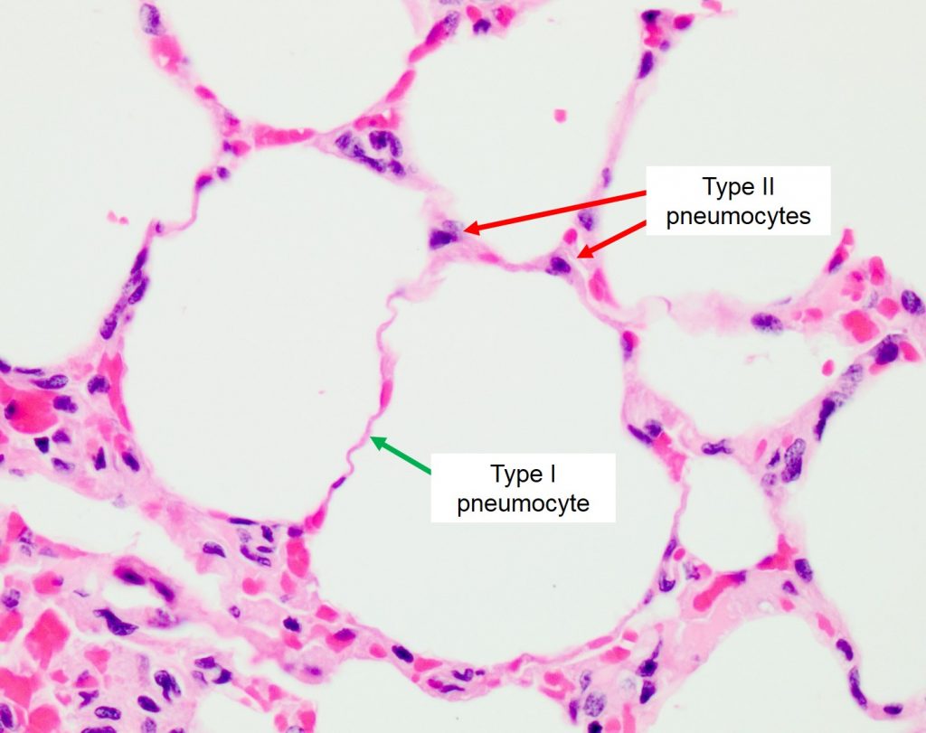
Alveoli (singular: alveolus) are the site of gas exchange in the lung. Alveoli are thin-walled, sac-like structures lined by a single layer of flattened squamous epithelial cells: type I pneumocytes. This extremely thin nature of the type I pneumocytes facilitates gas exchange across their surface. The alveolar wall, or septum, consists of capillaries and minimal connective tissue support. As such, alveolar septal capillaries are almost in direct apposition with the type I pneumocyte. The type I pneumocyte and capillary lumen are separated only by the basement membrane of the type I pneumocytes, minimal or absent septal connective tissue, the basement membrane of the capillary endothelium, and the endothelial cells themselves. This provides an extremely narrow gap through which gases can diffuse, providing for efficient exchange of oxygen and carbon dioxide between alveolar spaces and capillaries.
In addition to the type I pneumocytes, alveoli also contain several additional cell types. Type II pneumocytes are cuboidal epithelial cells frequently residing in the corners of alveolar spaces. Type II pneumocytes are responsible for the secretion of surfactant. Surfactant is a fluid composed of phospholipids and proteins that coats the surface of alveolar spaces to reduce surface tension, allowing for alveoli to expand and remain open. In addition, type II pneumocytes play a crucial role in tissue repair in the lung. Following damage to type I pneumocytes, type II pneumocytes proliferate and differentiate into type I pneumocytes, thereby restoring alveolar structure.
Finally, alveolar spaces contain a resident population of macrophages, alveolar macrophages. Normally, these macrophages are located in close apposition to type I pneumocytes, and can be mistaken for type II pneumocytes. These macrophages, which are relatively low in number, readily phagocytize debris within alveoli, which can include surfactant, edema fluid, red blood cells, and pathogens (e.g. bacteria). Low numbers of macrophages are also normally present within the alveolar septal interstitium.
