Chapter 12: Male Reproductive System
Male tubular genitalia
These structures are derived from the caudal remnants of embryonic structures called the mesonephric ducts (Wolffian ducts). They function to transport spermatozoa towards the urethra.
The structures that are derived from the mesonephric ducts include:
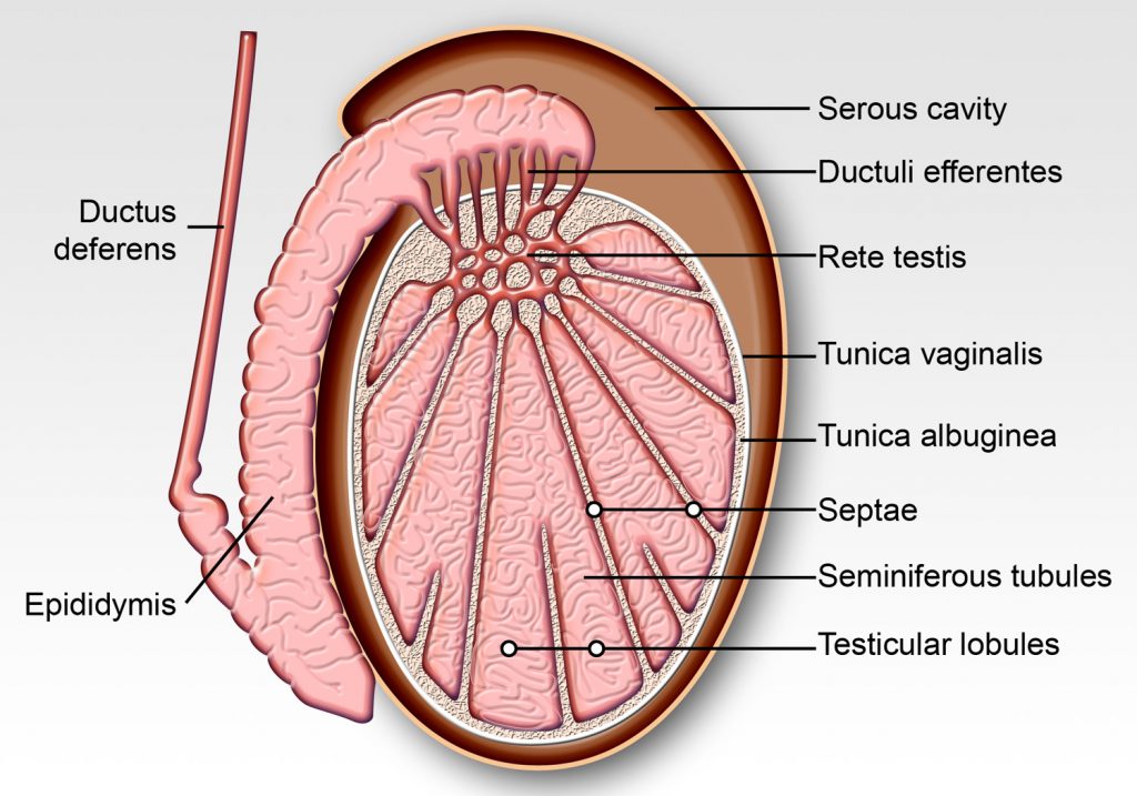
- Efferent ductules – These structures connect the rete testes to the ductus epididymis. They consist of 6-20 coiled tubules lined by ciliated columnar epithelium which assist in the movement of spermatozoa.
- Ductus epididymus – This coiled tube functions to store spermatozoa. Some maturation of spermatozoa also takes place here. The epididymus and its associated connective tissue and muscle are divided into the head (where the efferent ductules terminate), the body and the tail. The tail is continuous with the ductus deferens. The mucosa of the epididymus is lined by ciliated pseudostratified columnar epithelium. Under the lamina propria lies a thin band of smooth muscle (muscularis mucosa) which is slightly thicker at the tail. The submucosa of this structure is continuous with the connective tissue of the tunica albuginea.
- Ductus (vas) deferens – This structure is lined by pseudostratified columnar epithelium. In contrast to the previous tubular structures, the lamina propria lies on top of a thick smooth muscle tunica muscularis which is divided into inner and outer layers. The ductus deferens contains an outer serosa.