Chapter 12: Male Reproductive System
Development and structure of the gonads and tubular genitalia
Gonadal development
The first step in gonadal development takes place when the primordial germ cells migrate from the allantois (portion of the placenta) to the genital ridge, the structure that will become the undifferentiated gonad. The gonadal ridges are protuberances within the coelomic cavity of the developing fetus. The structures of the primitive gonad that are not composed of primordial germs cells (i.e. somatic gonadal cells) are derived from local mesenchymal cells, coelomic epithelium and cells derived from mesonephric tubules. Ultimately Sex determination of the undifferentiated gonad is dependent on the presence of the sex determining region Y (SRY) gene found on the short arm of the Y chromosome. Therefore without the Y chromosome and SRY gene, differentiation to a female gonad will take place.
Development of tubular genitalia
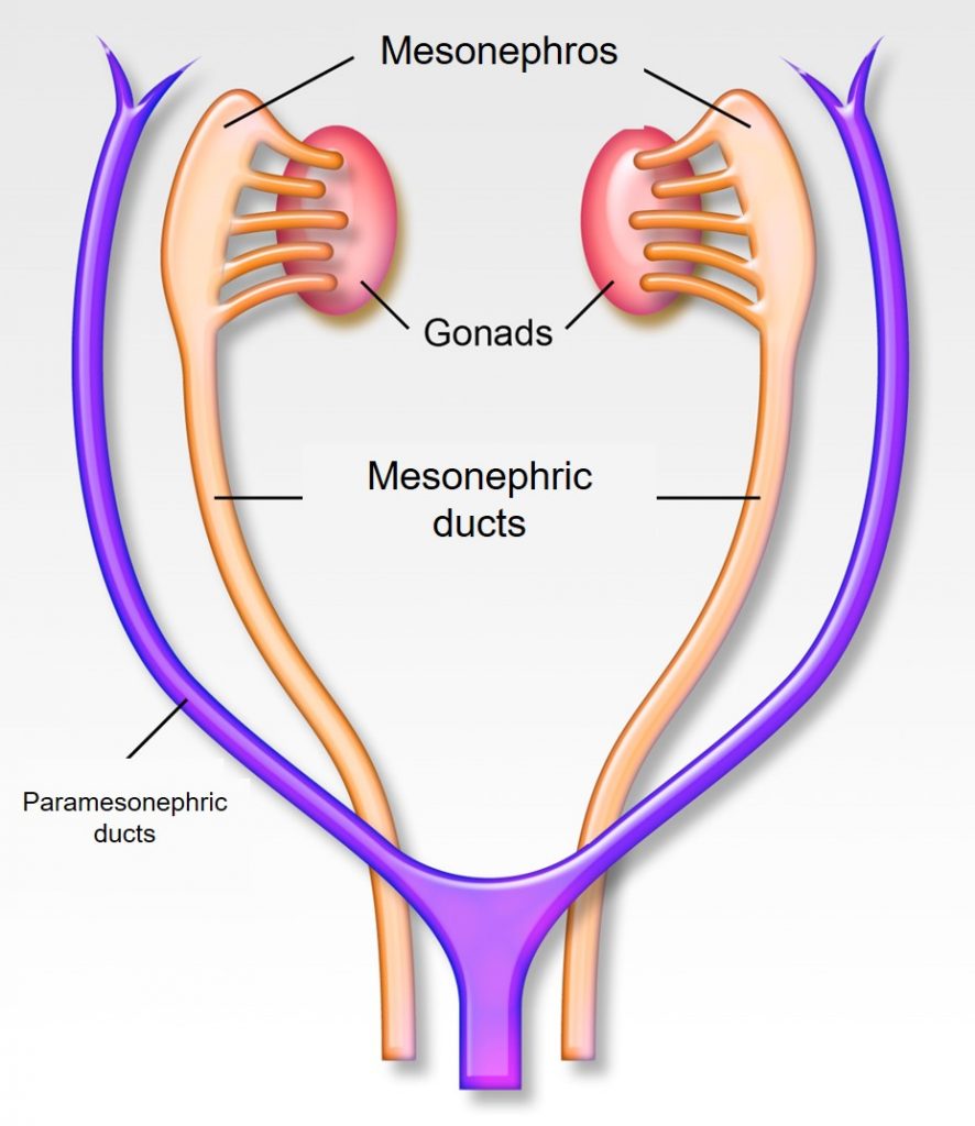
The formation of male tubular genitalia is directed by the hormones secreted by the developing testes. The secretion of testosterone from the Leydig cells and paramesonephric inhibitory hormone from the Sertoli cells induce the differentiation of the Wolffian body (mesonephros) and the Wolffian ducts (mesonephric ducts). Paramesonephric inhibitory hormone inhibits the development of the paramesonephric ducts (Muellarian ducts). In the absence of developing testes and the presence of estrogens, the paramesonephric ducts develop into the uterus, uterine tubes and cranial vagina. Estrogen also stimulates the development of female external genitalia, the caudal vagina, the vestibule and the clitoris (more in chapter 13).

