Chapter 14: The Eye
The Lens
The lens is a biconvex transparent structure and is the second refracting unit of the eye. It lies posterior to the iris and is suspended from the ciliary body by the zonular fibers. It also has a posterior attachment to the anterior vitreous face where it lies in a depression of the vitreous, the patellar fossa. It is a unique tissue in that it is avascular, transparent, lacks nerve supply, and has the highest concentration of protein in the body. Embryologically, the lens originates from the surface ectoderm that is induced to form the lens placode and invaginate by the advancing optic vesicle and cup.
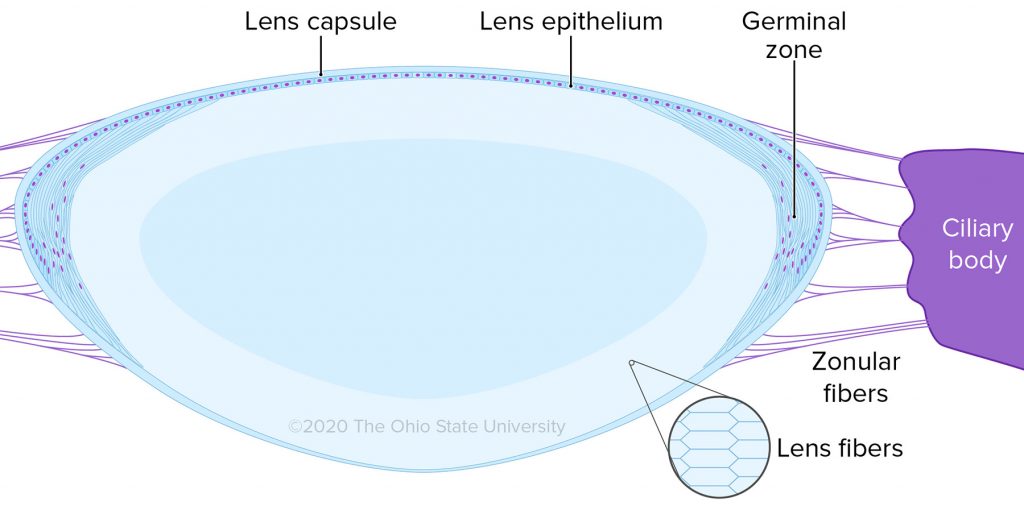
The lens is surrounded by a basement membrane, the lens capsule, which is secreted by the lens epithelial cells anteriorly and the cortical fibers posteriorly. The capsule is thickest anteriorly and thinnest at the posterior pole and continues to grow throughout life.
Beneath the lens capsule, anteriorly, is the lens epithelium. These form a monolayer of cuboidal cells whose apices face the cortical fibers with the basal portion of the cell adjacent to the lens capsule. The apical portion of the epithelial cells has terminal bars. Adjacent to the lens equator is the pre-equatorial or germinative zone where the lens epithelial cells replicate. It is these cells that are susceptible to radiation and toxic insult, resulting in cataract formation. The newly formed cells migrate equatorially where they elongate, differentiate into cortical fibers, are displaced inward, compressed, and loose their nuclei. This replicative process occurs throughout life. As these cortical fibers elongate anteriorly and posteriorly they attach at a line, the lens suture. In human, dogs, and cats these suture lines are in a Y-shape, upright anteriorly and inverted posteriorly, but this varies with other species. This results in a layered arrangement of the lens fibers with the oldest fibers in the center and the newest fibers surrounding them. On cross-section, the lens fibers are hexagonal and adjacent cells interdigitate with each other in the form of microplicae and by a ball-and-socket arrangement.

