Chapter 6: Cardiovascular System
Vascular tunics
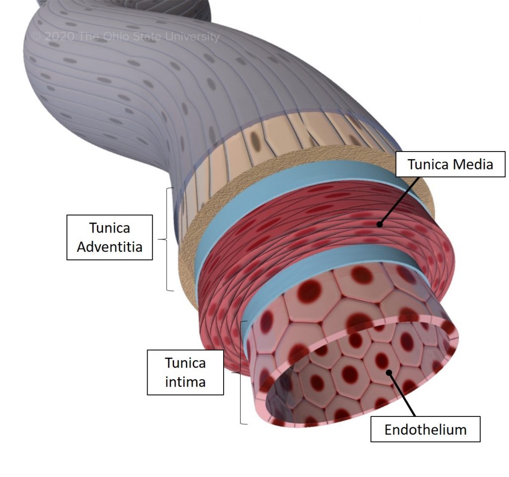
With the exception of capillaries and sinusoids, all larger vessels have the same three basic structural elements (tunics). These are the tunica intima (inner or luminal layer), tunica media (middle layer), and tunica adventitia (outer layer). Depending on the level of the vasculature, there are marked differences in the tunic width.

Endothelial cells are arguably the most important component of the tunica intima and form the primary barrier between blood and tissue. They are highly specialized cell type and cover the luminal surface of the tunica intima. Endothelial cells have a number of critical functions. During a resting phase, endothelial cells have important anti-clotting functions. They have anticoagulant effects (expression of heparin like molecules, production of thrombomodulin, tissue factor pathway inhibitor), inhibit platelet aggregation (ADPase, prostacyclin), and promote fibrinolysis (synthesis of tissue type plasminogen activator). Under certain pathologic states, endothelium contributes to a prothrombotic state by activating/aggregating platelets (ie. platelet activating factor, von Willebrand’s factor), inhibiting fibrinolysis (plasminogen activator inhibitors), and producing procoagulant factors (ie. tissue factor). Other functions include:
- Synthesis of collagen and proteoglycan for basement membrane maintenance (connective tissue interface)
- Modulation of blood flow by secretion and metabolism of vasoactive mediators (ie. nitric oxide, endothelin, angiotensin I)
- Degradation of catecholamines
- Expression of surface molecules that facilitate adhesion, rolling, and diapedesis of inflammatory cells
- Production of growth hormones (ie. FGF, PDGF)
- Production of molecules that mediate the acute inflammatory reaction (ie. IL-1, 6, 8)
- Antigen presentation
Endothelial cells can be identified by their location. They are the flattened, fusiform cells lining the lumen of vessels. Von Willebrand’s factor can be contained in Weibel-Palade bodies that might be apparent under electron microscopy. Other ultrastructural features include cytoplasmic vesicles that reflect phagocytic and pinocytotic activity. Highly specialized endothelial modifications can be present depending on the organ and location (see discussion on capillary structure and function). Supporting the vascular endothelium, and forming the remainder of the tunica intima, is a basement membrane and a subendothelial layer. The subendothelial layer contains collagen, elastic fibers and proteoglycan; smooth muscle cells, fibroblasts and myofibroblasts are also present. Collagen, contained within the subendothelium, is a particularly important stimulus for platelet activation and adhesion. Fibronectin helps to stabilize cell-cell and cell-substrate interactions.
The tunica media is the middle portion of the vessel wall. It contains extracellular matrix (elastic laminae, ground substance), and circumferentially arranged smooth muscle cells. The internal and external elastic laminae delineate the inner and outer limits of the tunica media, respectively. These elastic laminae become harder to identify in closer proximity to the microcirculation. In histologic sections of arteries, arteries contract and the internal elastic lamina has extensive tortuous folds. In arteries, the tunica media is extremely thick and is the primary constituent of the vessel wall. The venous supply, which operates at considerably lower pressure, has an extremely thin tunica media. Unlike arteries, the internal elastic lamina is only prominent in medium and large caliber veins.
The tunica adventitia is the outermost layer of the vessel wall. It is composed of a loose layer of connective tissue, containing vasa vasorum, a vascular bed designed to perfuse the vessel wall itself. The tunica adventitia is particularly prominent in large caliber veins (such as the caudal vena cava), where it may also contain bundles of loosely arranged smooth muscle responsible for maintaining venous tone. The adventitia of large veins is richly innervated by nerves and also contains lymphatic vessels.
