Chapter 8: Gastrointestinal System
Small intestine
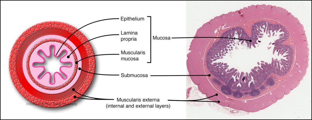
The small intestine of most domestic species is quite similar in function and histology. Structural and functional differences in specific regions of the small intestine impart differential functional capabilities to these segments. The small intestine is divided into three distinct segments, from oral to aboral: duodenum, jejunum, and ileum. The major functions of the small intestine are digestion, secretion, and absorption. The small intestinal mucosa has several anatomic adaptations that serve to create an immense surface area with which to digest and absorb nutrients. These include the plicae circulares (intestinal folds), villi, and microvilli.
The small intestinal mucosa is characterized by numerous, regularly distributed luminal papillary projections called villi. Villi are lined by columnar epithelial cells, enterocytes. Enterocytes have apical microvilli (brush border). Although individual microvilli are indistinguishable, the brush border is apparent as a faintly staining, uniform ~1 µm layer along the superficial surface of enterocytes. Enterocytes function mainly in digestion and absorption. The enterocytes are interspersed with goblet cells, columnar epithelial cells with abundant, poorly staining cytoplasm, representing mucin. The density of goblet cells is generally more abundant in more distal segments of the intestine.
The intestinal villi are contiguous with glands located at the base of villi: the crypts of Lieberkuhn, or intestinal crypts. The crypts contain the mitotically active population of intestinal epithelial stem cells. Mitotic figures are commonly seen in cells of the crypts. Within the crypts, epithelial stem cells divide and push upward (luminally), further differentiating into enterocytes or goblet cells. In this way, the small intestinal villi are similar to a production conveyor belt –intestinal epithelial cells are generated in the crypts and progressively migrate superficially along the villi towards the luminal surface where, at the tips of villi, the epithelial cells are sloughed into the lumen. This process occurs continually and promotes a high rate of enterocyte replacement/turnover.
In addition to enterocytes and goblet cells, the small intestine contains low numbers of accessory cells. In some species such as the horse, the crypts contain low numbers of cells, Paneth cells, that contain abundant eosinophilic cytoplasmic granules. These granules contain antimicrobial molecules important in gut innate immunity. Finally, low numbers of enteroendocrine cells are interspersed within the crypts. These enteroendocrine cells produce hormones that may include somatostatin, cholecystokinin, and secretin. Enteroendocrine cells are not readily apparent in routine H&E sections.
The lamina propria of the small intestine extends into and forms the core of small intestinal villi. The villous lamina propria is composed primarily of loose collagenous tissue, but contains a number of important structures and cells. The villous lamina propria is rich in both capillaries and lymphatics that help transport nutrients absorbed by enterocytes across the luminal surface. The small intestinal lamina propria also contains low numbers of immune cells, including lymphocytes and plasma cells, and small numbers of lymphocytes are regularly located within the villous epithelium.

Microscopic anatomic features
Although much of the previously discussed features of the small intestine apply to the duodenum, jejunum, and ileum, distinguishing microscopic anatomic features of the duodenum and ileum are detailed below.
Duodenum
The gastric pylorus empties into the lumen of the duodenum. The duodenal submucosa contains extensive tubuloacinar glands, Brunner’s glands, that are lined by tall columnar epithelial cells with mucin-rich, poorly-staining cytoplasm. The Brunner’s glands communicate with the lumen of the crypts of Lieberkuhn. The secretions of the Brunner’s glands are alkaline and help to neutralize the acidic digesta received from the stomach.
The pancreatic duct and common bile duct insert into the wall of the duodenum and communicate with the duodenal lumen.
Ileum
The ileal mucosa contains large numbers of organized lymphoid tissue (lymphoid follicles), termed Peyer’s patches. The Peyer’s patches serve as both a primary and secondary lymphoid organ. Peyer’s patches are critical components of the GALT.
